
28-04-2026 20:07
 Lothar Krieglsteiner
... on twig in the air at standing Ceratonia siliq
04-05-2026 18:13
 Stephen Martin Mifsud
ID request for what seems to be a true aquatic fun
04-05-2026 16:39
Stephen Martin Mifsud
ID request: This specimen was collected in Malta o
04-05-2026 09:50
 Castillo Joseba
Me mandan el material seco de Galicia,(España) re
02-05-2026 12:42
Alain BRISSARDBonjour à tousJeuidi 30 avril dernier on m'a remi
02-05-2026 13:06
Pauline. PennaBonjour Please can someone help me with this id
01-05-2026 22:45
 Thierry Blondelle
Bonjour à tous, Une récolte sur bouse séchée d
14-04-2026 05:32
Ethan CrensonHi all, A few weeks back a friend pointed out som
28-04-2026 20:33
Vitus SchäfftleinHello, I found Trochila ilicina on Ilex aquifoliu
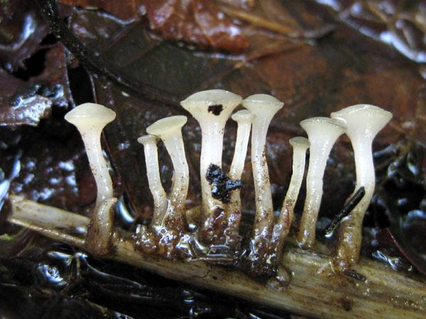
??Dear everyone.
This discomycete, frequently found in Japan, seems to be identical to Cudoniella clavus in macroscopic, microscopic, and ecological characters.
According to Dennis, etc., ascus pore of C. clavus is non-amyloid, but this ascus pore turns blue by IKI, more clearly with KOH pre-treatment. (I have no microphoto)
Is it true C. clavus?
Masanori Kutsuna

In Huhtinen 1985: 511 (mycoflora Poste-de-la-Baleine?) the reaction is clearly blue after KOH. Also In my collections I saw repeatedly a distinct blue reaction in IKI (Hymenoscyphus-type).
However, at least in one (HB 1000, from Black Forest) ?I noted IKI-. This explains why authors like Dennis, Breitenbach & Kränzlin or Gamundi 1998: 114? likewise found inamyloid asci.
Zotto?
Thank you for the answer.
Let me ask you a question.
If so, what is the difference between Hymenoscyhus and Cudoniella?
Regards,
Masanori Kutsuna
Zotto
Regards,
Kutsuna