
10-06-2026 21:16
 François Freléchoux
Bonsoir,Le dernier du jour, en attendant votre avi
11-06-2026 19:01
 William Slosse
Hello all,In an attempt to make a culture of a sus
11-06-2026 19:03
 Nicolas VAN VOOREN
Chers membres d'Ascofrance,Le site sera placé en

09-06-2026 18:32
Camille MertensSur morceau de roseau immergé 0,5 - 0,7 mm de dia
10-06-2026 12:54
Steve ClementsBonjour encore, Pouvez-vous m'aider, s'il vous pl
10-06-2026 21:07
François Freléchoux
Toutes les tiges de gentianes jaunes de l'an pass�
10-06-2026 13:41
François Freléchoux
Bonjour à nouveau, Voici une trouvaille d'hier.
10-06-2026 11:53
Steve ClementsBonjour, This disco is abundant on dead stems of

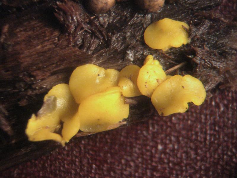
Hello, forum!
There is Bisporella collected on Populus tremula.
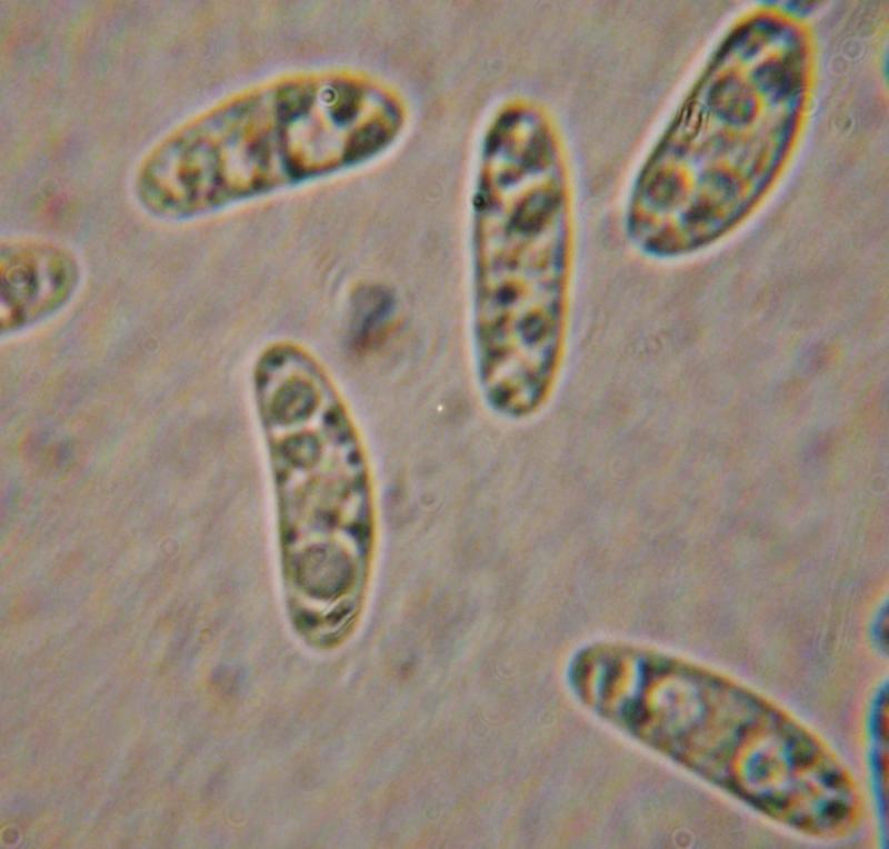
Micro:
In statu vivo:spores ellipsoid, 14,9-19,3-3,6-5,5 um, with 2-3 large and many small guttules.
Paraphyses guttulate (photo is of low quality and I can attach the drawing if needed)
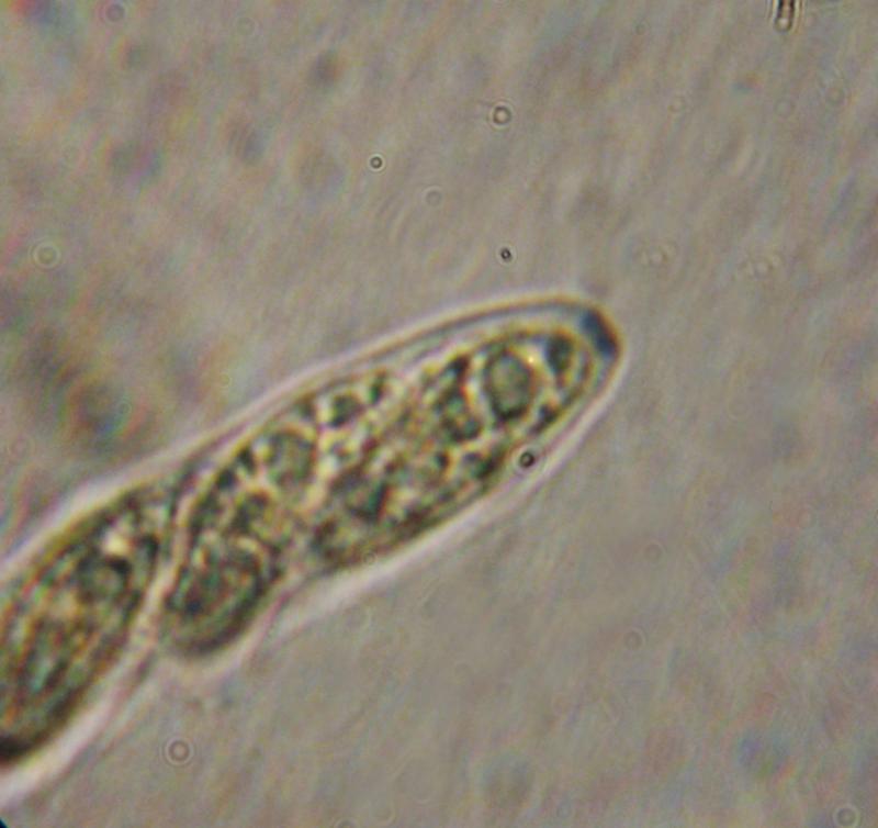
Asci with croziers, 8-spored, blued in IKI (see photo)
Is it B. citrina? If it is so, why paraphyses are guttulate?
Sincerely, Irina


the paraphysis is strange, it should contain a homogeneous content in the upper part. Otherwise I also think B. citrina. A section is always good, excipulum should be gelatinized and the hyphae with a almost vertical orientation.
Zotto

Yes, but be careful, maybe you ovelooked very elongated (with homogenous content) vacuolar bodies in apical cells (the microphoto is very blur) because sometimes they can be of +/- whole cell length. Considering small "guttules" they could be isolated/concentrated yellow carotenoid pigment - the situation I observed in some other ascomycetes with carotenoid pigments where pigment can be separated from VB's and/or LB's. In such cases VB's and LB's are often highly refractive and hyaline or nearly so while pigment are decidedly yellow to orange to red and low refractive if at all...
At least asci are clearly of Calycina type :-) !!
Cheers,
Neven
Thank you, Zotto and Neven!
Really, in the upper part paraphyses were not-guttulate (probably, gelatinous, but I cannot say it exactly). In the lower part paraphyses contained many small rounded guttules (it's not seen from photo but I saw them very clearly).
Sincerely,
Irina