
28-04-2026 20:33
Vitus SchäfftleinHello, I found Trochila ilicina on Ilex aquifoliu
28-04-2026 22:51
 Bernard CLESSE
Bonsoir à toutes et tous,Pourriez-vous m'aider à

28-04-2026 21:50
 Pablo Sandoval
Hola a todos,Espero se encuentren bien. Hace mucho
27-04-2026 18:05
 Lothar Krieglsteiner
... still attached at standing tree. The green con
28-04-2026 20:07
Lothar Krieglsteiner
... on twig in the air at standing Ceratonia siliq
27-04-2026 20:52
Lothar Krieglsteiner
Found on hanging tiwg of Olea europaea in dried-ou
27-04-2026 18:48
Tony MoverleyCollected 23rd April 2026, Norfolk, EnglandSwarms
27-04-2026 17:41
Lothar Krieglsteiner
.. Algarve, same leaf than the last post. The con
27-04-2026 17:16
Lothar Krieglsteiner
.. Algarve, moist lying.The conidiomata look like
Pithya vulgaris info
Raúl Tena Lahoz,
05-06-2011 20:55
 Hi to all!
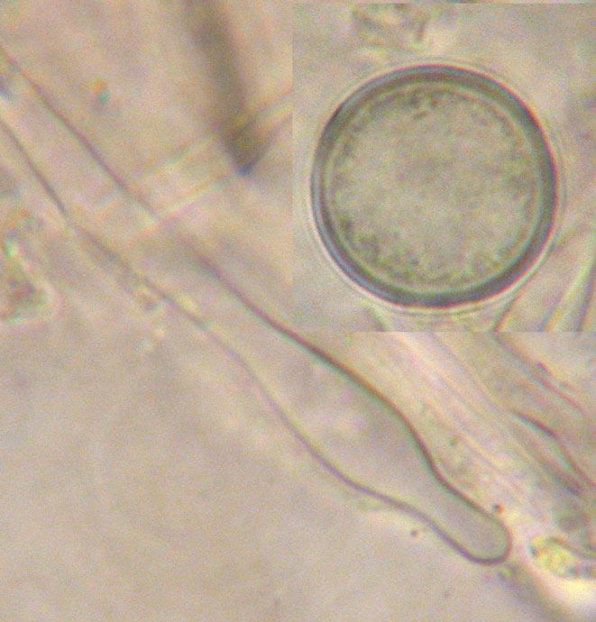
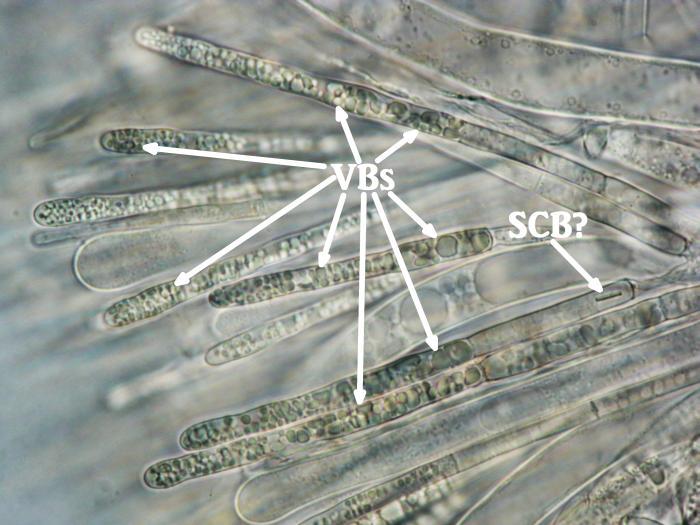
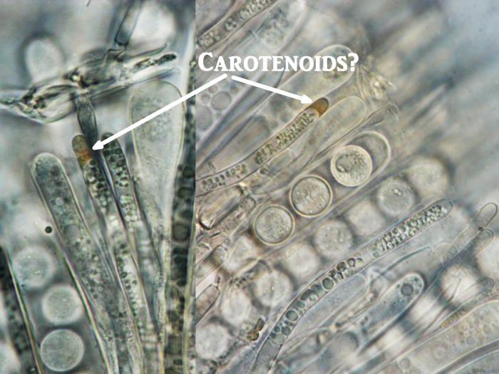
I´m testing the changes (bravo!) and wanted to ask for vital microphotos or drawings of Pythia vulgaris. Specially about paraphyses (VBs and carotenoids), and ectal excipulum. Also about nuclei information when spores are fully mature. I know they have four nuclei (Zotto), but finally they duplicate more times?
Thanks in advance!
Raúl
Hans-Otto Baral,
05-06-2011 23:32

Re : Pithya vulgaris info
Hi Raul
I only have this photo of Pithya vulgaris (by JP Dechaume). I do not know of VBs in the paraphyses.
Zotto
I only have this photo of Pithya vulgaris (by JP Dechaume). I do not know of VBs in the paraphyses.
Zotto
Raúl Tena Lahoz,
06-06-2011 13:27
Re : Pithya vulgaris info
Thanks Zotto
I was wondering how carotenoids appear inside paraphyses of Pithya vulgaris. In Pithya cupressi I suppose that first the VBs develop and then appear the carotenoids. But I´m not sure if this occurs in this way. It is a type of oxidation? Somebody talks about this processes? Maybe Arpin?
Raúl
I was wondering how carotenoids appear inside paraphyses of Pithya vulgaris. In Pithya cupressi I suppose that first the VBs develop and then appear the carotenoids. But I´m not sure if this occurs in this way. It is a type of oxidation? Somebody talks about this processes? Maybe Arpin?
Raúl
Raúl Tena Lahoz,
06-06-2011 13:28
Re : Pithya vulgaris info
Carotenoids
Raúl Tena Lahoz,
06-06-2011 13:31
Re : Pithya vulgaris info
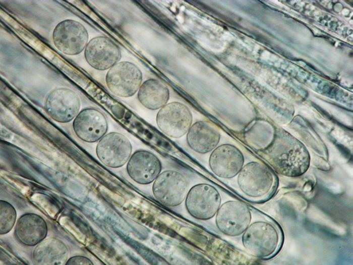
Also seen 4 nuclei inside spores, but if you don´t use software to join photos it is very difficult to show it with only an image. You can see at least three nuclei/nucleoli in some of the spores.
Raúl
Raúl
Nicolas VAN VOOREN,
06-06-2011 18:27

Re : Pithya vulgaris info
Hi Raul.
On a collection made in March 2007, on dead branches of Abies alba, I have written "paraphyses with guttules and yellowish-orange content... ectal excipulum of textura angularis, with cells 15-20 µm, hyaline". No information about nuclei.
On a collection made in March 2007, on dead branches of Abies alba, I have written "paraphyses with guttules and yellowish-orange content... ectal excipulum of textura angularis, with cells 15-20 µm, hyaline". No information about nuclei.
Raúl Tena Lahoz,
06-06-2011 19:35
Re : Pithya vulgaris info
Merci beaucoup Nicolas!
Hans-Otto Baral,
07-06-2011 18:26
Re : Pithya vulgaris info
I do not think that carotenoids and VBs are correlated. VBs may turn reddish-brownish through oxidation, but the result is never something like carotenoids. Carotenoids are coloured in the living state.
I am surprised that you found VBs in Pithya. It seems I never saw them, but I admit I have little documentation. - Oh, now I see I have the VBs in my drawing of Pithya cupressina, HB 2426.JPG?
Zotto
I am surprised that you found VBs in Pithya. It seems I never saw them, but I admit I have little documentation. - Oh, now I see I have the VBs in my drawing of Pithya cupressina, HB 2426.JPG?
Zotto
Raúl Tena Lahoz,
07-06-2011 20:02
Re : Pithya vulgaris info
Yes, I saw your drawing Zotto.
Then, the colours of Pithya are because the presence of carotenoids or not?
Another question: carotenoids are evident from the first stages of development of apothecia (in any genera that have them)?
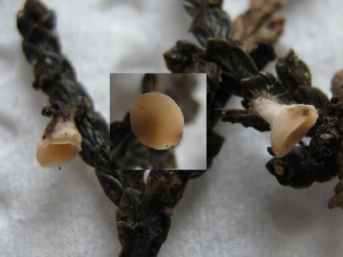
I´m used to see nearly white Pithya cupressi in the first stages of development and when they are covered by the Cupressus/Juniperus leaves. I attach the macro of the apothecia to which the microphotos belong.
Raúl
Then, the colours of Pithya are because the presence of carotenoids or not?
Another question: carotenoids are evident from the first stages of development of apothecia (in any genera that have them)?
I´m used to see nearly white Pithya cupressi in the first stages of development and when they are covered by the Cupressus/Juniperus leaves. I attach the macro of the apothecia to which the microphotos belong.
Raúl
Hans-Otto Baral,
07-06-2011 21:23
Re : Pithya vulgaris info
Yes, surely the colour is only because of the carotenoids. As you can see from externally, the colour is present already when very young. Yes, I also know this pale form. Development of carotenoids is probably supported by light, but I am unaware of pale apos getting bright-coloured with age, if the light conditions are not changed.
The new forum looks very nice, but when typing the answer, I can only see Raul's first mail, not his last to which I wish to answer....
Zotto
The new forum looks very nice, but when typing the answer, I can only see Raul's first mail, not his last to which I wish to answer....
Zotto
Raúl Tena Lahoz,
08-06-2011 10:03
Re : Pithya vulgaris info
Well, then I have problems to find the carotenoids in these pale forms. I don´t know where to look for them.
In the previous photo, where I asked for carotenoids in paraphyses, I signaled pigments that in my opinion are the responsables of the apothecia colour. Because I saw them in paraphyses where the VBs where scanty or starting to dissolve, then I thought they were related to the VBs. Do you think they are not carotenoids? Maybe just an oxidation of the VBs?
Me too, I would prefer to see the hole evolution of the dicussion. By now I just open a new window.
Raúl
Hans-Otto Baral,
08-06-2011 11:01
Re : Pithya vulgaris info
My impression is that the pigment on your photos is outside the vacuoles which would point to carotenoids. Usually you can stain carotinoids with IKI (in greenish), but mainly when they are strongly coloured.
Zotto
Zotto
Raúl Tena Lahoz,
08-06-2011 14:53
Re : Pithya vulgaris info
Also carotenoids should resist KOH?
Stip Helleman,
09-06-2011 00:11

Re : Pithya vulgaris info
Hi Raúl,
indeed it is strange i cant see the whole discussion while reponding, yesterday i thougt there was work on the site because i could not repond. Today i found out Internet explorer did not sign me in automatically.
The carotene in paraphyses are not in all species evident when young, in Neodasyscypha and Proliferodiscus the guttules appear more greenish when young so the Hymenium looks more black
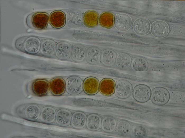
for the Pithia vulgaris i found in januari but did not make any notes about the excipulum or paraphyses but i made a IKI test for the spores to see if there were any glycogenes. The sporewall turned blue on the outside and red on the inside so there seems to be some glycogene accumilation on the inside of the sporewall. For nuclei I did not look but judging my photo i see only two
cheers,
Stip
indeed it is strange i cant see the whole discussion while reponding, yesterday i thougt there was work on the site because i could not repond. Today i found out Internet explorer did not sign me in automatically.
The carotene in paraphyses are not in all species evident when young, in Neodasyscypha and Proliferodiscus the guttules appear more greenish when young so the Hymenium looks more black
for the Pithia vulgaris i found in januari but did not make any notes about the excipulum or paraphyses but i made a IKI test for the spores to see if there were any glycogenes. The sporewall turned blue on the outside and red on the inside so there seems to be some glycogene accumilation on the inside of the sporewall. For nuclei I did not look but judging my photo i see only two
cheers,
Stip
Raúl Tena Lahoz,
09-06-2011 12:21
Re : Pithya vulgaris info
Thanks Stip
Did you take data about living spores measured in water? I´m also interested in that info. I see that Nicolas talks about (13) 14-16.
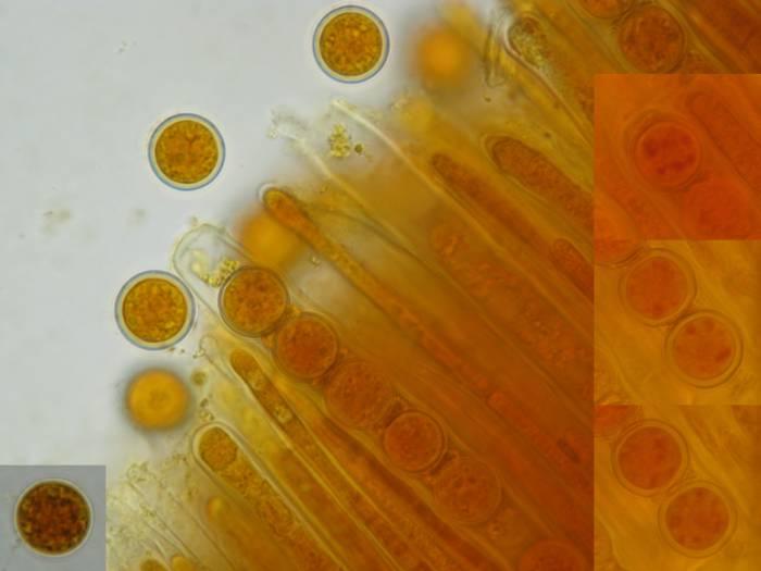
I noticed glycogen in Pithya cupressi, see pics. Also seen this kind of blue aureole. But, at least in my case, I think it is because chromatic aberrationsof the ocular/objective. Maybe it is enhanced due to the characteristics of the outer wall in this genus. I add also a picture in IKI + KOH with different focus and you can see with one focus the aureole but not if you change the focus. In those spores where the outer wall is more separated, the blue aureole is enhanced, maybe the bigger space and the light passing through IKI gives this blue appearance (aberration)... I don´t know.
Raúl
Did you take data about living spores measured in water? I´m also interested in that info. I see that Nicolas talks about (13) 14-16.
I noticed glycogen in Pithya cupressi, see pics. Also seen this kind of blue aureole. But, at least in my case, I think it is because chromatic aberrationsof the ocular/objective. Maybe it is enhanced due to the characteristics of the outer wall in this genus. I add also a picture in IKI + KOH with different focus and you can see with one focus the aureole but not if you change the focus. In those spores where the outer wall is more separated, the blue aureole is enhanced, maybe the bigger space and the light passing through IKI gives this blue appearance (aberration)... I don´t know.
Raúl
Hans-Otto Baral,
09-06-2011 12:31
Re : Pithya vulgaris info
I also need to log in every day....
Carotenoids do not disappear in KOH, yes, but when VBs get oxidated, the colour may be also quite resistent to KOH.
The bluish and reddish colour around the spore wall I also think is an optical effect, like in the rainbow. Not due to microscope optics, only because of differences in the density of spore wall and water arond.
Zotto
Stip Helleman,
10-06-2011 00:39
Re : Pithya vulgaris info
Hi Raúl & Zotto,
This color reaction is too obvious i think for a optical effect. In fact it remembers me of the basidiospores of Leucocoprinus capillipes (the outer wall, inside they have one large Glycogene body) i tested with IKI and they are said to be achromatic, next time i will test Cressyl bleu on the spores. I am sorry but i did not make any measurements of anything.
Cheers,
Stip
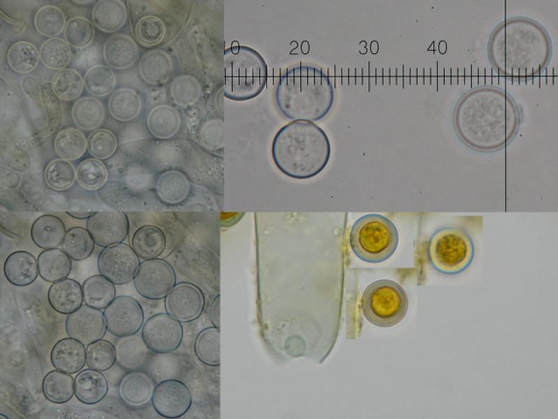
Raúl Tena Lahoz,
12-06-2011 16:47
Re : Pithya vulgaris info
Thanks Zotto & Stip
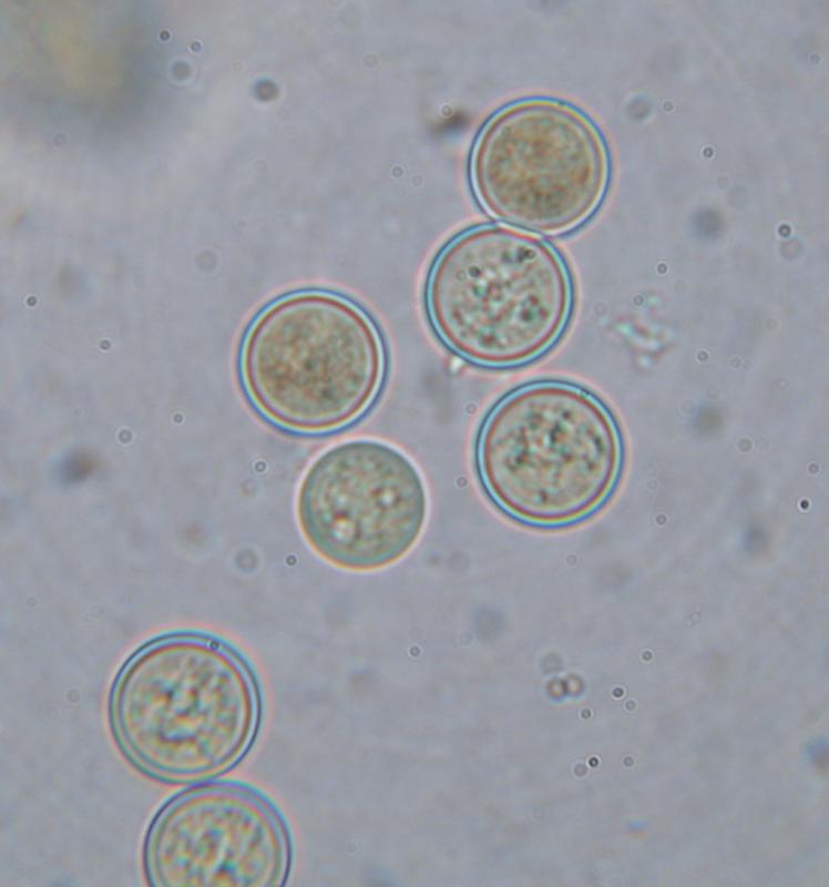
Here I attach a photo Plicaria leiocarpa spores. All in water except those of the right hand at the bottom. Because I see the blue aureole also in those in water, I think It has to be an optical efect, at least in my microscope.
Raúl
Here I attach a photo Plicaria leiocarpa spores. All in water except those of the right hand at the bottom. Because I see the blue aureole also in those in water, I think It has to be an optical efect, at least in my microscope.
Raúl
Stip Helleman,
13-06-2011 01:01
Re : Pithya vulgaris info
Hi Raúl & Zotto,
it seems you both are right, it is an alternation of the light spectrum. And i get curiuos now what causes that, most likely it is not the optics when it is seen in 3 different microscopes. Next time i will do some more extensive tests, or it could be the sporeshape perhaps
cheers
Stip
it seems you both are right, it is an alternation of the light spectrum. And i get curiuos now what causes that, most likely it is not the optics when it is seen in 3 different microscopes. Next time i will do some more extensive tests, or it could be the sporeshape perhaps
cheers
Stip