18-11-2024 00:06
• Macro and habitat seem hyaloscyphoid.• Hairs

15-11-2024 20:08
• Macro and habitat seem mollisiod.• Mollisia

14-11-2024 12:06
 carl van den broeck
On November 8th I found very small orange discs st

14-11-2024 15:31
 Bernard CLESSE
Bonjour à toutes et tous,Que pensez-vous de ce Sc

14-11-2024 04:18
 Götz Palfner
Dear community, is this Nemania carbonacea? Micros

14-11-2024 00:34
• Apothecia with predominantly yellow or brown h

11-11-2024 23:17
• Macro and habitat suggest Hyaloscyphaceae s.l.

12-11-2024 16:43
Ethan CrensonHello all, This weekend a friend found these dark

13-11-2024 08:01
 Stephen Martin
I am revising some old material again and I have t
"Tapesia" with crystals puzzle
Chris Yeates,
24-05-2015 19:33
 Bonsoir tous
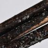
I recently collected a discomycete with abundant pale apothecia on dead stems of Phragmites, cut and moved away from the water. It had an abundant dark chocolate subiculum, and based on the shortish spore size in the past, and using Ellis and Ellis, I would have tentatively called this Mollisia (Tapesia) evilescens. However I note from Andreas Gminder's key that the type of that taxon has cells which are brownish all the way to the margin; in my fungus all except the basal attachment and cells immediately surrounding it were not pigmented. I also note from Andreas' key that this is one of the difficult areas of Mollisia studies.
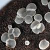
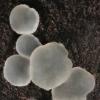
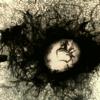
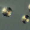
A particular feature of this fungus was the presence of globose bodies comprising numerous tiny acicular crystals. I wondered at first whether these were from an external source, but found them in all the several apothecia I studied. In addition I also could see refractive bodies within cells of the medulla base (see 7th image below). NB In the accompanying images the colours in the crystal bodies are due to polarised light, used to accentuate them, they were in themselves colourless.
Apothecia were white to pale grey, and up to 1mm diameter. No KOH reaction noted at either the macro- or micro- level.
Asci 8-spored, apex IKI deep blue, with croziers.
Ascospores 9-10.9 x 2.5-2.7µm.
Paraphyses, typical Mollisia-type with vacuolar contents.
Mollisia hydrophila is perhaps the closest species, but I note that that fungus has abundant crystals (not the case here); also as I understand it those crystals are of the familiar octohedral type (again not the case here). In addition that species should show a KOH+ reaction. Plus, that species has basal pads of dark hyphae, whereas this fungus had an extensive subiculum, encircling the Phragmites stems. While I appreciate that presence of a subiculum is not significant at the generic level, surely it has some taxonomic significance at the specific level?
Any suggestions to help ease my confusion would be welcome.
Cordialement
Chris
Hans-Otto Baral,
24-05-2015 20:59

Re : "Tapesia" with crystals puzzle
Hi Chris
I also find only M. hydrophila as the closest match, and agree that a widespread subiculum is not known there, nor is the absence of a yellow KOH-reaction. The shape of crystals I would not estimate as high, but they look indeed characteristic. In hydrophila they partly also form aggregations too, but not like yours.
I understand that you mean that "all excipular cells except for the basal ones are not pigmented".
I think that Andreas has not enough studied these monocot-inhabiting species to provide a comprehensive key. But I assume that mine leads likewise not to a satisfying solution.
The VBs in globose cells should originate from cortical excipular cells, not medullary. Did you see this in a section or only a squash mount?
Zotto
Chris Yeates,
25-05-2015 03:20

Re : "Tapesia" with crystals puzzle
Thanks for the comments Zotto
you are correct - I meant to say " all except the basal attachment and cells immediately surrounding it were not pigmented" and have now corrected that error in my original post.
The structures within the globose cells appeared to me to be crystalline, not VB's, but I shall endeavour to cut some sections (not one of my skills); you are right - the image is from a squash. I shall also re-check the presence or absence of a KOH reaction. That subiculum does however remain problematical . . .
LG
Chris
Hans-Otto Baral,
25-05-2015 08:23

Re : "Tapesia" with crystals puzzle
I seem to have been mislead by the turquoise colour of these drops: I thought you stained in CRB, but now I think it is because of polarised light? If so that would plead for crystals. But these drops are inside the cells, aren't they?
