
10-06-2026 21:16
 François Freléchoux
Bonsoir,Le dernier du jour, en attendant votre avi
11-06-2026 19:01
 William Slosse
Hello all,In an attempt to make a culture of a sus
11-06-2026 19:03
 Nicolas VAN VOOREN
Chers membres d'Ascofrance,Le site sera placé en

09-06-2026 18:32
Camille MertensSur morceau de roseau immergé 0,5 - 0,7 mm de dia
10-06-2026 12:54
Steve ClementsBonjour encore, Pouvez-vous m'aider, s'il vous pl
10-06-2026 21:07
François Freléchoux
Toutes les tiges de gentianes jaunes de l'an pass�
10-06-2026 13:41
François Freléchoux
Bonjour à nouveau, Voici une trouvaille d'hier.
10-06-2026 11:53
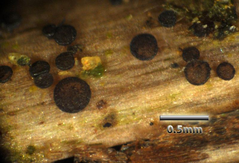
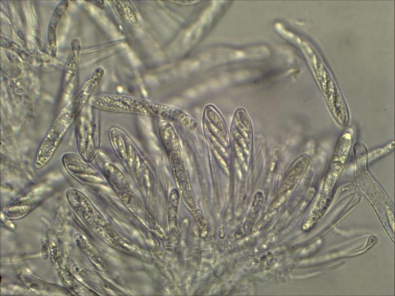
Steve ClementsBonjour, This disco is abundant on dead stems of
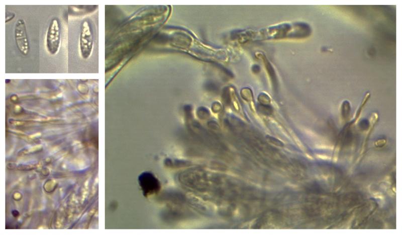
 Collected on a dead attached rachis of Dryopteris dilatata at 255 metres above sea level in the Yorkshire Pennines, England. The encrusted excipular margins and strongly dextrinoid paraphysis tips are very distinctive, but I have drawn a blank: this seems not to be among any of the Helotiales recorded on Dryopteris in the UK.
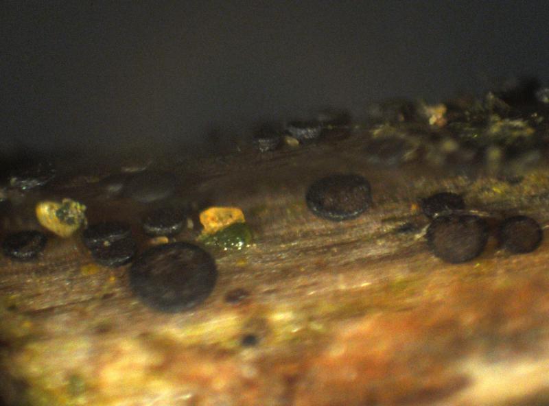
Apothecia flat with slightly raised rims, sessile, up to circa 400µm diameter; dull brown with darker margins.
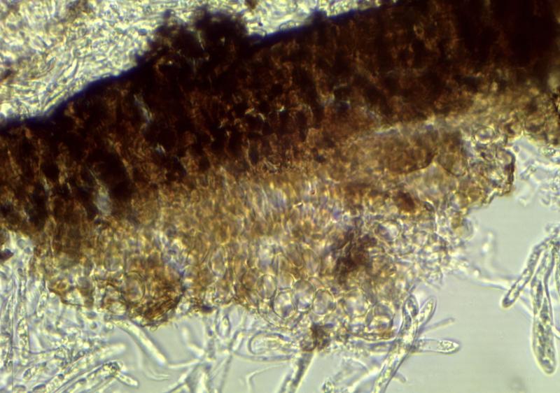
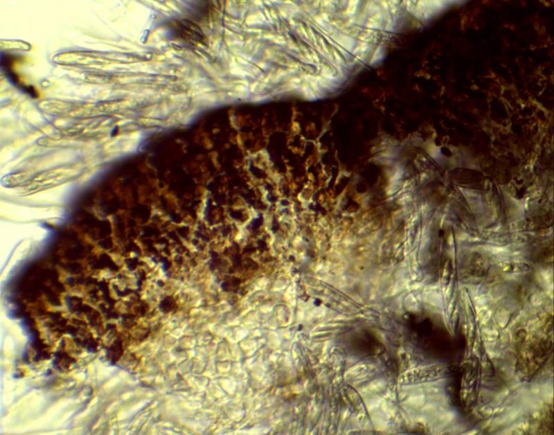
Excipulum from basal hyaline textura globulosa/prismatica to a margin heavily encrusted with dark brown material.
Asci 8-spored 48-60 x 6-8µm, pore blueing in IKI;ascospores elliptic-fusiform 9.75-10.5 x 3µm.
Paraphyses with rather thick-walled apices which are dextrinoid and variously clavate/obpyriform/capitate in shape.
Any suggestions very much appreciated .
Chris


the paraphyses are not thick-walled but they contain a refractive vacuole in the inflated apex. You did not see them in water? It would be intersting whether they were hyaline or perhaps yellow.
This is clearly a "Crustomollisia", a genus which can either be talen as a synonym of Calycellina, or treated as Calloriella or Micropeziza p.pt. I presently prefer to say Calloriella.
I have here species on herbaceous stems (C. umbrinella = Allophylaria soederholmii), Quercus leaves (c. castanea = Crustom. roburnea), Cyperaceae/Poaceae (Pseudonaevia caricina ?= Actinoscypha muelleri; Micropeziza cornea/karstenii), and some others, but none on ferns.
You see from this that it is a rather confused though very interesting group, with these brown clods of exudate on the exterior.
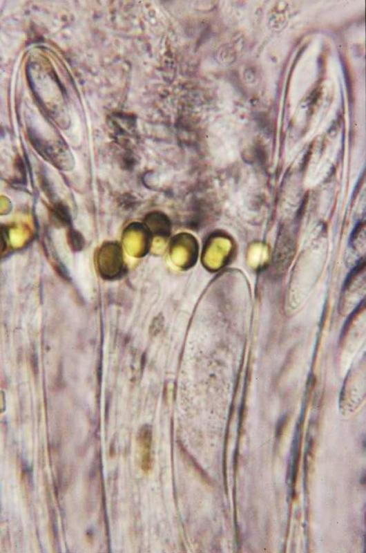
I attach here a photo of C. castanea with yellow VBs in the paraphyses.
If there is anything in the literature that fits to your fungus I will see in my database.
Zotto
If you could get some free spores (I assume it is not easy) then I could better see the contents. I think the spores are aseptate and contain larger and smaller oil drops.
The VBs you can only see in the living paraphyses. f you see them, you can try also Cresyl Blue for staining them. If there are any spores around, please check if the surface stains lilac in CRB.
Zotto
I am keeping it damp for a day or so to ensure ripe spores, I am pretty certain they are non-septate though; I don't have cresyl blue, but Derek Shafer has put me on to a UK supplier who sells it in dry form as Brilliant Cresyl Blue; some places sell it as a solution - the formula being (this I understand is sold for diagnostic blood testing) 1% Cresyl Blue and 0.85% sodium chloride in water?
is the NaCl actually needed?
thanks
Chris
No NaCl necessary, this is, was you say, only for cells that do not have a cell wall. In fungi you would provoke a certain shrinkage of the cell because you take away the turgor.
Zotto
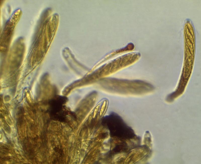
Yes, it has proved difficult to get free spores; see the accompanying image, the spores looked very similar to those in the asci being almost full of oil drops - I did however see a single spore (pictured - hidden beneath "PREV"!) which looks unequivocably septate; the VB's in the paraphysis tips are indeed pale yellowish (rather as in your image) . . . . all these are of living material in water
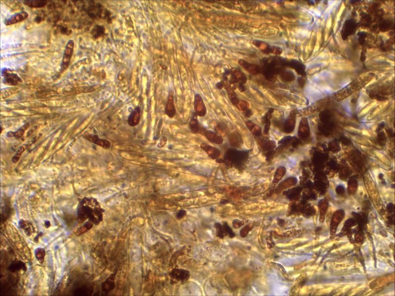
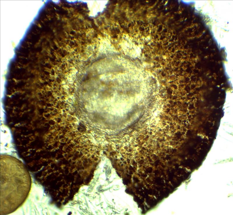
the excipulum is almost entirely covered in exudate as can be seen in the other photograph; this does not match at all well with a description of Pezizella roburnea in TBMS 67(3) page 481:
"margin formed of compacted slightly pigmented pseudo-parenchymatous cells"; I do not have access to Velenovsky's original description. Also the habitat is very different - am I correct in thinking there are not many ascomycete species which occur on both Pteridophyte and Spermatophyte plants? I know a few fern rusts alternate with Gymnosperms but that is a different matter
best wishes
Chris
I attach here the paper of Svrcek where you see the brown patsches on the excipulum. Don't know what Hawksworth & Sivanesan saw (on Castanea leaves).
The high lipid content in your spores clearly excludes Calloriella castanea = Crustomollisia roburnea. I have a collection on Vaccinium stems from lapland which resembles your fungus also in the spores, but it was not very wel developed and remained unidentified. Spores were ca. *14-15 x 5-5.5 µm?, so yours are smaller.
Did you measure the free spores? They have a sheath which is typical of Calloriella. The septum is probably only present at an overmature stage.
Maybe it is an undescribed species, I found nothing on ferns in my database.
Zotto
 Svrcek-1987-Crustomollisia-Fuscoscypha-0001.pdf