
30-07-2025 18:06
Stefan JakobssonOn a decorticated twig of Alnus incana on moist so


13-06-2025 09:41
 Josep Torres
Hello.A cerebriform ascomycete sprouting scattered
13-06-2025 16:34
 Andgelo Mombert
Bonjour,Un petit discomycète qui me résiste. Il
21-07-2025 19:22
Ethan CrensonHello all, Here is an Orbilia found by a friend
14-07-2025 11:20
 Michel Hairaud
Bonjour, Voici une espèce de (?) Hyaloscyphace
18-07-2025 23:03
Josep Torres
Hello.Fruitings between 51 and 130 microns in tota
Orbilia dryadum?
Stefan Jakobsson,
30-07-2025 18:06
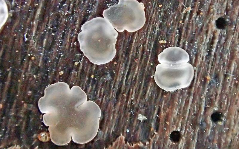
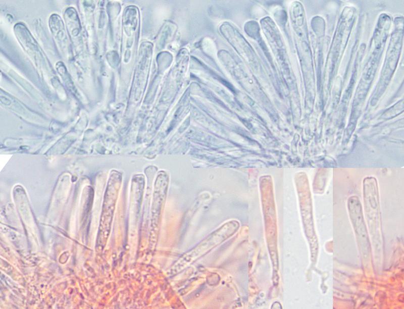
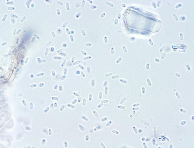
On a decorticated twig of Alnus incana on moist soil there was a small group of Orbilas with apos 0.5-1.5 mm. The spores are on avg 3.1 × 1.3 µm and Q = 2.46. The asci are on avg. 29.0 × 3.2 µm.
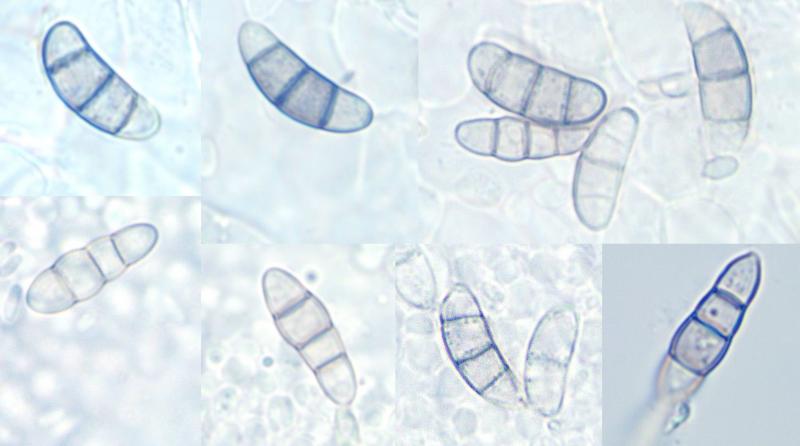
Most details fit nicely to Orbilia dryadum but the conidia don't, they are slighly bent and the size is 12.8 - 16.2 × 4.6 - 5.6 µm. Perhaps they do not belong to the Orbilia even thogh they were found under apothecia? Southern Finland.
What should I think about this one?
Hans-Otto Baral,
31-07-2025 09:03

Re : Orbilia dryadum?
This fits perfect indeed. Any brown conidia found in association with Orbiliomycetes are aliens and belong to other fungi.
Stefan Jakobsson,
31-07-2025 13:15
Re : Orbilia dryadum?
Thank you for the confirmation!