11-05-2026 12:32
 Bernard CLESSE
Pourriez-vous m'aider à identifier cette héloti
13-05-2026 15:26
 François Freléchoux
Bonjour,Voici une récolte faite il y a quelques j
12-05-2026 15:41
 Nicolas VAN VOOREN
Dear Ascolovers, especially interested in Pezizale
13-05-2026 12:05
 Thierry Blondelle
Bonjour à tous,J'aimerais avoir confirmation de c
10-05-2026 23:17
 Andreas Gminder
Hello,today we found in a moist steep decidous for
28-04-2026 20:07
 Lothar Krieglsteiner
... on twig in the air at standing Ceratonia siliq
27-04-2026 20:52
Lothar Krieglsteiner
Found on hanging tiwg of Olea europaea in dried-ou
11-05-2026 20:22
Lothar Krieglsteiner
on attached twig of standing Ficus caricaquite uns
29-04-2026 10:44
Lothar Krieglsteiner
growing at moist, drying-out soil at the side of a
Lophodermium on Pinus pumila
Mathias Hass,
08-04-2025 11:22

Hi,
I have some trouble with this Lophodermium. I suppose there are 3 possible species...but which one? I hope some can help.
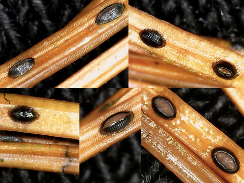
Found in March in large numbers on attached needles of Pinus pumila (a 5-needle species) from a botanical garden, Denmark. P. pumila is not a naturally occurring tree in Denmark. Apotecia in conspicuously regular intervals along the needles, very few transverse black lines, on one occasion a brown line.
Apotecia: about 0,63 mm (0,46 mm - 0,81 mm) long (n=10), ellipsoid. No clear black line surrounding the apotecia. Lips look to me greyish. On one occasion seen in bright sunlight the lips appeared bluish. I do not feel sure about the colour, but red they are not.
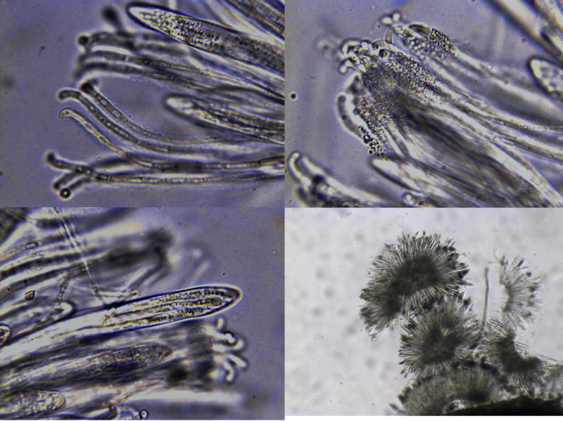
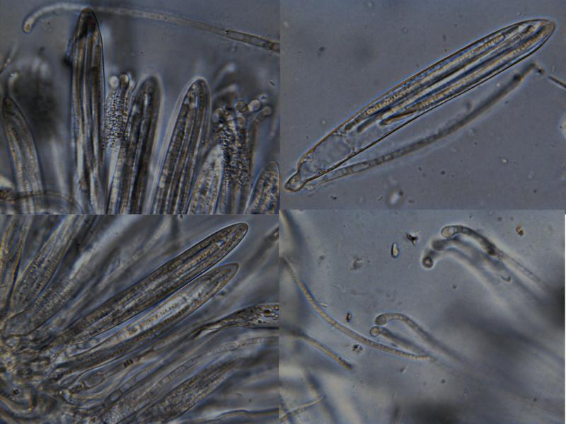
Asci: when mature > 100 my long, up to about 130 x 15 my, typically 116x12 my, 8 spores arranged in parallel.
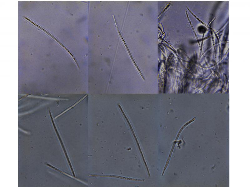
Ascospores: Direct after collection (few spores): 83,5 (73,9-93,0) my x 1,9 (1,8-2,1) my, Q=43, n=10. Surrounded by a 2,4 my thick gelatinous sheet. No appendages seen. After incubation wet and at RT for a week: 73,7 (63,3-87,6) x 2,3 (2,0-2,8) my, Q=32 , n=10. The spores seem to become significantly shorter but wider (and paraphyses get swollen tips).
Paraphyses: cylindrical, 2,5 my wide, somewhat bent tips but not swollen. The tips have a large number of diffracting small droplets which makes them appear brownish, at least in relatively immature apotecia. After incubation pararaphyses have swollen tips, up to 4 my broad.
Asci: when mature > 100 my long, up to about 130 x 15 my, typically 116x12 my, 8 spores arranged in parallel.
Ascospores: Direct after collection (few spores): 83,5 (73,9-93,0) my x 1,9 (1,8-2,1) my, Q=43, n=10. Surrounded by a 2,4 my thick gelatinous sheet. No appendages seen. After incubation wet and at RT for a week: 73,7 (63,3-87,6) x 2,3 (2,0-2,8) my, Q=32 , n=10. The spores seem to become significantly shorter but wider (and paraphyses get swollen tips).
Paraphyses: cylindrical, 2,5 my wide, somewhat bent tips but not swollen. The tips have a large number of diffracting small droplets which makes them appear brownish, at least in relatively immature apotecia. After incubation pararaphyses have swollen tips, up to 4 my broad.
Kind Regards
Mathias