

22-04-2026 20:54
 Enrique Rubio
Hi to everybody.This Pyrenopeziza grew in moist le
24-04-2026 03:16
 David Chapados
Found while looking at something else from wood in
22-04-2026 01:06
 Richard VALERI
Bonjour à tous.Je vous présente cette Nectria s.
22-04-2026 20:17
 Marian Jagers
Is anyone familiar with the Hyphomycetes genus Pse

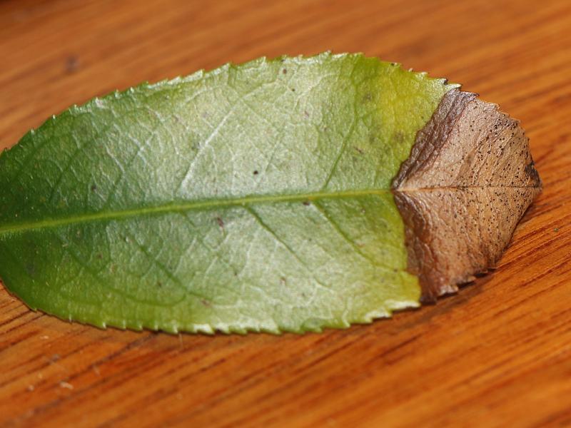
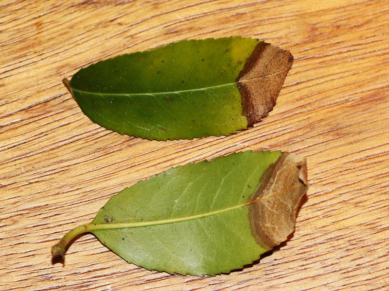
 On 6 february 2025 i stumbled upon a leafspot on Prunus lusitanica in Aerdenhout (The Netherlands). Can someone confirm that it is indeed Coleophoma prunicola? or maybe something completly different?
 For more photo's see: https://waarneming.nl/observation/338565267/
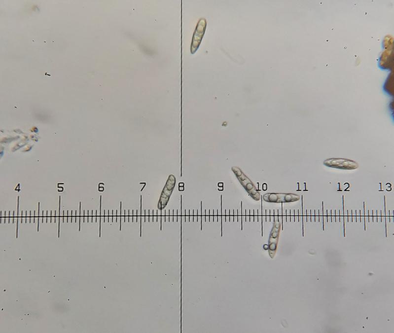
Conidiomate pycnidia, ca. 150- 200 µm diameter (N=10).
Conidia holoblastic, hyaline, aseptate, cylindrical, apex obtuse, base truncate, thin-walled, smooth, guttulate with several large guttules.
Conidia 20,8 - 23,4 - 26 ?m × 4-5,2 ?m (N=25)
400 X mafnification: 1 div. = 2,6 ?m
Following the Key for Coleophoma in the article: 'Reinstatement of Coleonaema for Coleophoma oleae and notes on Coleophoma' it should be Coleophoma prunicola
(Duan, J.X., Wu, W.P. and Liu, X.Z. (2007). Reinstatement of Coleonaema for Coleophoma oleae and notes on Coleophoma. Fungal Diversity 26: 187-204.) https://www.researchgate.net/publication/237440307_Reinstatement_of_Coleonaema_for_Coleophoma_oleae_and_notes_on_Coleophoma
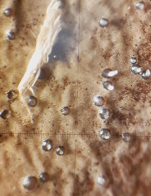
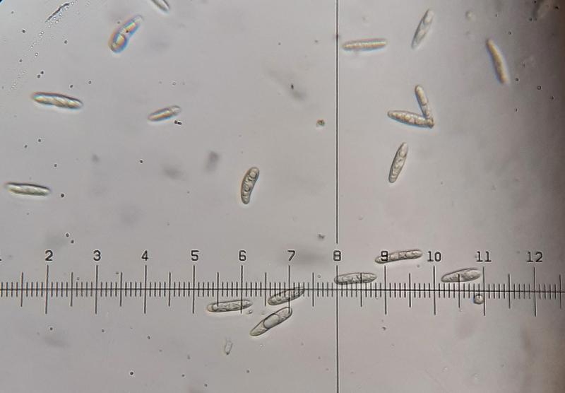

To know this you should see the conidiogenous cells and compare them with those in the article.
Best wishes
Angel
Thanks for your comment. I can try another time to see the conidiogenous cells and compare them with those in the article. I already tried once but failed to see the conidiogenous cells sadly.
Kind regards,
Jorian