
09-02-2026 14:46
Anna KlosGoedemiddag, Op donderdag 5 februari vonden we ti

02-02-2026 21:46
Margot en Geert VullingsOn a barkless poplar branch, we found hairy discs

07-02-2026 20:30
 Robin Isaksson
Hi!Anyone that have this one and can sen it to me?

25-01-2026 23:23
Hello! I found this species that resembles Delitsc

05-02-2026 15:07
Found on a fallen needle of Pinus halepensis, diam

05-02-2026 06:43
Stefan BlaserHello everybody, Any help on this one would be mu

18-08-2025 15:07
 Lothar Krieglsteiner
.. 20.7.25, in subarctic habital. The liverwort i
Hyaloscypha on dead leaf of Myrica gale.
François Bartholomeeusen,
09-09-2024 16:40
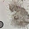
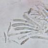
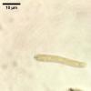
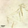
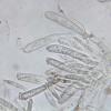
Thorough inspection yielded only one fruit body. I did not have a second attempt at microscopy, but given the specific shape of the spores and the fact that the asci contain only four spores, I identify this asco as Hyaloscypha lachnobrachya.
Would anyone be so kind to confirm that!
Thank you,
François Bartholomeeusen
Diameter fruit body: 155 µm
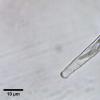
Asci: 34 x 5 µm; IKI +; Croziers +
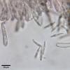
Spore: (15.3) 15.4 - 16.1 (16.6) × (1.7) 1.9 - 2.5 (2.6) µm; Me = 15.8 × 2.2 µm; Qe = 7.2
Hans-Otto Baral,
09-09-2024 16:57

Re : Hyaloscypha on dead leaf of Myrica gale.
Hi Francois
you are almost right, it is close to Calycellina lachnobrachya, which is no Hyaloscypha at all but a Pezizellaceae. Remnants of the VBs are visible in you not well intact specimens.
The species is polyphagous, but what me wonders is the high oil content in the spores.
Do you have my old paper on this group?
Baral, H.O. (1989). Beiträge zur Taxonomie der Discomyceten II. Die Calycellina-Arten mit 4sporigen Asci. – Beitr. Kenntn. Pilze Mitteleuropas 5: 209–236.
Zotto
François Bartholomeeusen,
09-09-2024 18:31
Re : Hyaloscypha on dead leaf of Myrica gale.
Thank you Zotto for your quick answer!
Unfortunately, I do not have the publication you cited, could you send it to me?
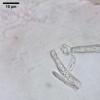
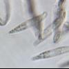
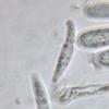
I added two detailed photos of the spores. I forgot to mention that the fruit body was completely white.
Unfortunately, I do not have the publication you cited, could you send it to me?
I added two detailed photos of the spores. I forgot to mention that the fruit body was completely white.
Best regards,
Hans-Otto Baral,
09-09-2024 21:14

Re : Hyaloscypha on dead leaf of Myrica gale.
I send it to you.
François Bartholomeeusen,
19-09-2024 20:01
Re : Hyaloscypha on dead leaf of Myrica gale.
Dear forum members,
I would like to finalise my item of 9 September on the forum. Mr Baral's splendid publications provided a lot of information but some uncertainties remain.
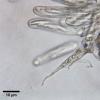
Calycellina is evident by the presence of a brown basal ring and the presence of light refracting vacuoles in excipulum, hairs and parphyses.
The high percentage of oil droplets in the spores (> 50%), the width of the spores and the absence of a stipe leaves two candidates namely C. flaveola and C. ulmariae. The substrate and texture of the extal excipulum complicate the choice. The only fruiting body I found was on a fallen leaf and the textura is difficult to determine. Perhaps the textura is 'oblita' after all? What are your thoughts?
Many thanks,
François
Hans-Otto Baral,
19-09-2024 21:11

Re : Hyaloscypha on dead leaf of Myrica gale.
I would say the hairs are apically acute :-) Because acute cell ends do not exist. C- ulmariae is strictly on Filipendula as I remember. Likewise C. flaveola is only on Pteridium.
I have now divided Calycellina in two folders, one with 4-spored asci. There I created a folder myricariae and pit your find as well as one by Michel Hairaud. This seems to me the same species, with a rather high lipid content too. From Michel I have no spore size to compare.
Oddly enough, both of you did not succeed with a macrophoto.
My guess is a new species. In my database is nothing on Myrica with such a spore size.

 AF-Is-Calycellina-ulmariae-een-oplossing-0001.docx