
12-06-2026 14:50
 François Freléchoux
Bonjour, Voici la brève description d'une Mollis
10-06-2026 21:16
François Freléchoux
Bonsoir,Le dernier du jour, en attendant votre avi
11-06-2026 19:01
 William Slosse
Hello all,In an attempt to make a culture of a sus
11-06-2026 19:03
 Nicolas VAN VOOREN
Chers membres d'Ascofrance,Le site sera placé en

09-06-2026 18:32
Camille MertensSur morceau de roseau immergé 0,5 - 0,7 mm de dia
10-06-2026 12:54
Steve ClementsBonjour encore, Pouvez-vous m'aider, s'il vous pl
10-06-2026 21:07
François Freléchoux
Toutes les tiges de gentianes jaunes de l'an pass�
10-06-2026 13:41
François Freléchoux
Bonjour à nouveau, Voici une trouvaille d'hier.
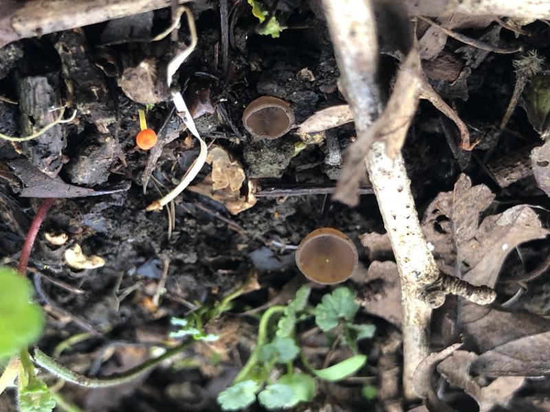
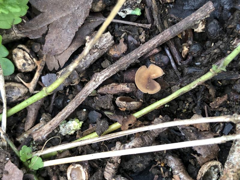
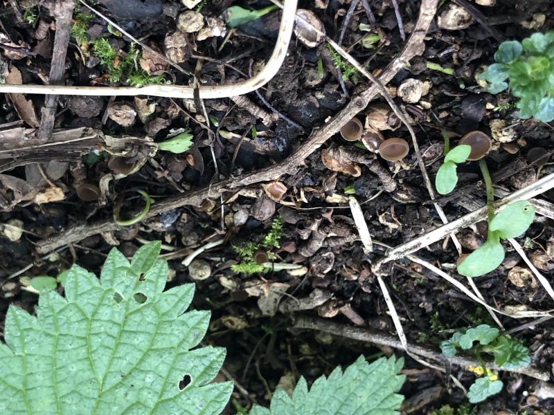
 It seems that this species is easily determined from the teleomorph by the habitat and macro, but I did some microscopy anyway.
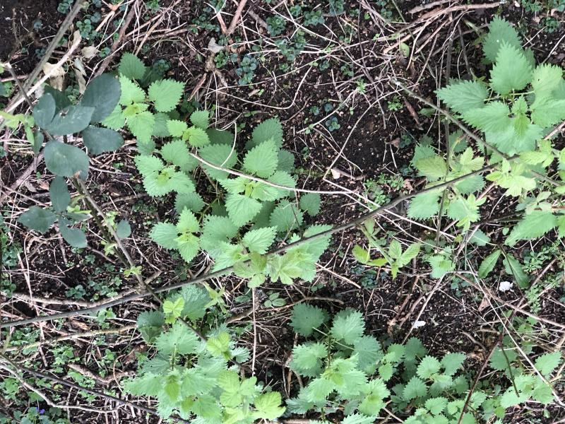
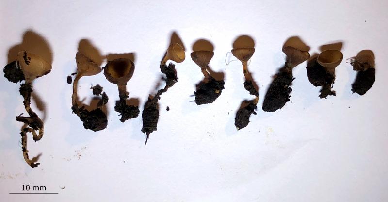
Habitat: On overwintered, stromatised haws (fruit) of Crataegus monogyna, in litter or buried, in a mature glade, on a small hill, South Downs, England, late March with the first leaves of the host.
Associates: Lots of nematodes in the soil around the haws, one basidioma of Mycena acicula on a small twig, often with some herbaceous ground cover.
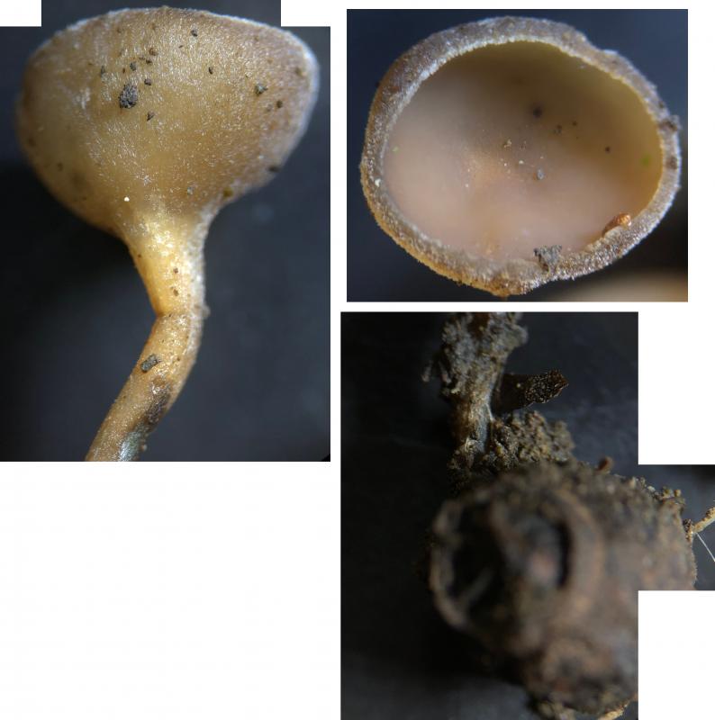
Apothecia: Measurements including full stipe length (3) 7-20 (30+) x 4-7 mm, n = 9, mean = 13 x 5 mm, Q = (0.8) 1.5-3.9 (4.2), mean Q = 2.3, goblet-shape, stipitate, cyathiform to cupulate and eventually +/- plane, deep to light brown, apparently darker with age, blackening when over mature, generally well-camouflaged amongst old upturned half-pits; receptacle and stipe with downy appearance; margin distinct, frosted-pruinose appearance, often in-rolled, with fringe of encrusted hairs/glassy processes?; disc concave, smooth appearance, more creamy colour; stipe 2-27+ x 1-1.5 mm, length depending on the depth that the haw is buried, when longer usually undulating more, appearance like the receptacle, often blackening near the base.
Storage and methods: Stored in a damp box for ~36 hours while still attached to haw, then stipe removed, cut in half, and slices taken from each side of the inside edge (not very effective as need more practise), mounted in water, some squashing later to separate the cells of the hymenium, IKI added to water mount, lots of bacteria in water mount.
Asci: Cylindrical, apex rounded to obtuse, croziers, IKI strongly bb – sclerotinia type, 1-1.5 seriate, one spore at apex, no inversely oriented spores observed.
- Living : ~130-190 x 9-12 um, pars sporifera ~20%, sub-biseriate when fully turgid, spores obliquely arranged, little to no apical thickening visible.
- Dead: ~7.5-9 um wide, pars sporifera > ~60%, uniseriate, apex more obtuse, distinct apical thickening when dead ~2.5-3 (4.8) um high.
Spores: Ellipsoid-obovate, often heteropolar with the apex rounded and the base more acute, occasionally more limoniform, rarely slightly curved, appearing binucleate with numerous medium-size shadowy LBs towards each pole and surrounding the nuclei, OCI 2-3, thick-walled, appear to have a perispore.
Measurements of mature vital spores in water mount: (11.4) 12.3-13.8 (14.1) × (6.3) 6.4-7.5 (7.6) µm, n = 21, mean = 12.9 × 7 µm, Q = (1.6) 1.7 - 2 (2.1), mean Q = 1.8.
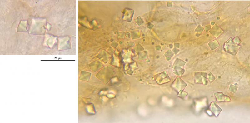
Exudate: Orange-red, in subhymenium and medullary excip., appears more patchy in ectal excip., some clumping on surface; rhomboid crystals of varying sizes, hyaline, often in groups, apparently on the ectal excip.; appears to be a hyaline exudate or glassy processes around the marginal hairs.
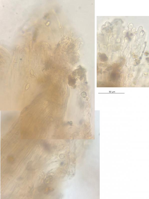
Paraphyses: Cylindrical, apices rounded, slightly inflated to slight tapering - 3.5-4 (5) um wide, multi-septate, appear to have large hyaline guttules, possibly sensitive to trauma.
Ectal excipulum: Thin layer of hyphae externally with short hyphal tips/hairs protruding, then textura prismatica-globosa.
Marginal hairs: Like paraphyses, apical cell more inflated and capitate, sometimes irregularly inflated, protruding above the hymenium.
Subhymenium and medullary excipulum: Difficult to distinguish, textura intricata, separated from ectal excip. by broad band of horizontal hyphae.


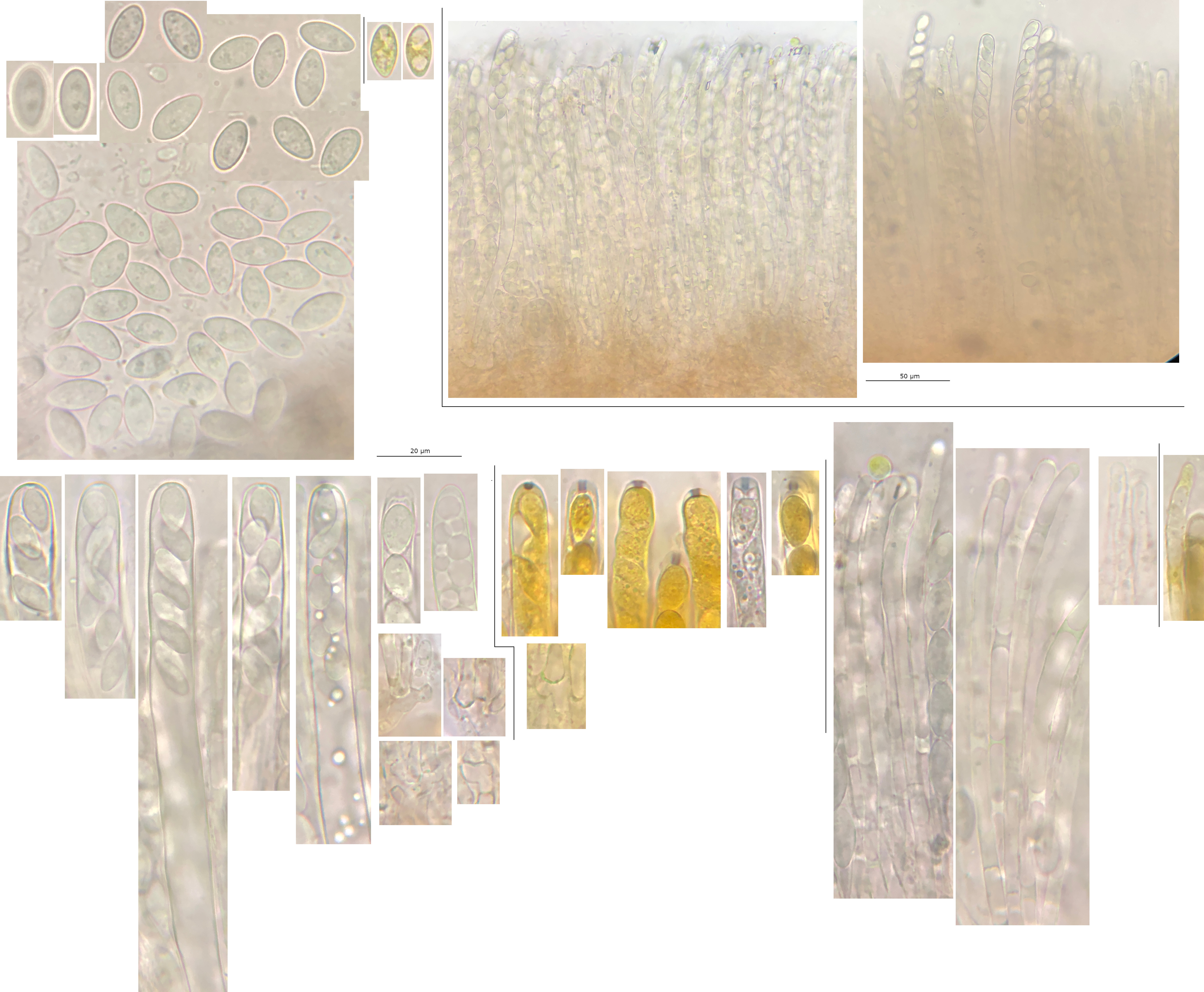 Hymenium-0008.jpeg
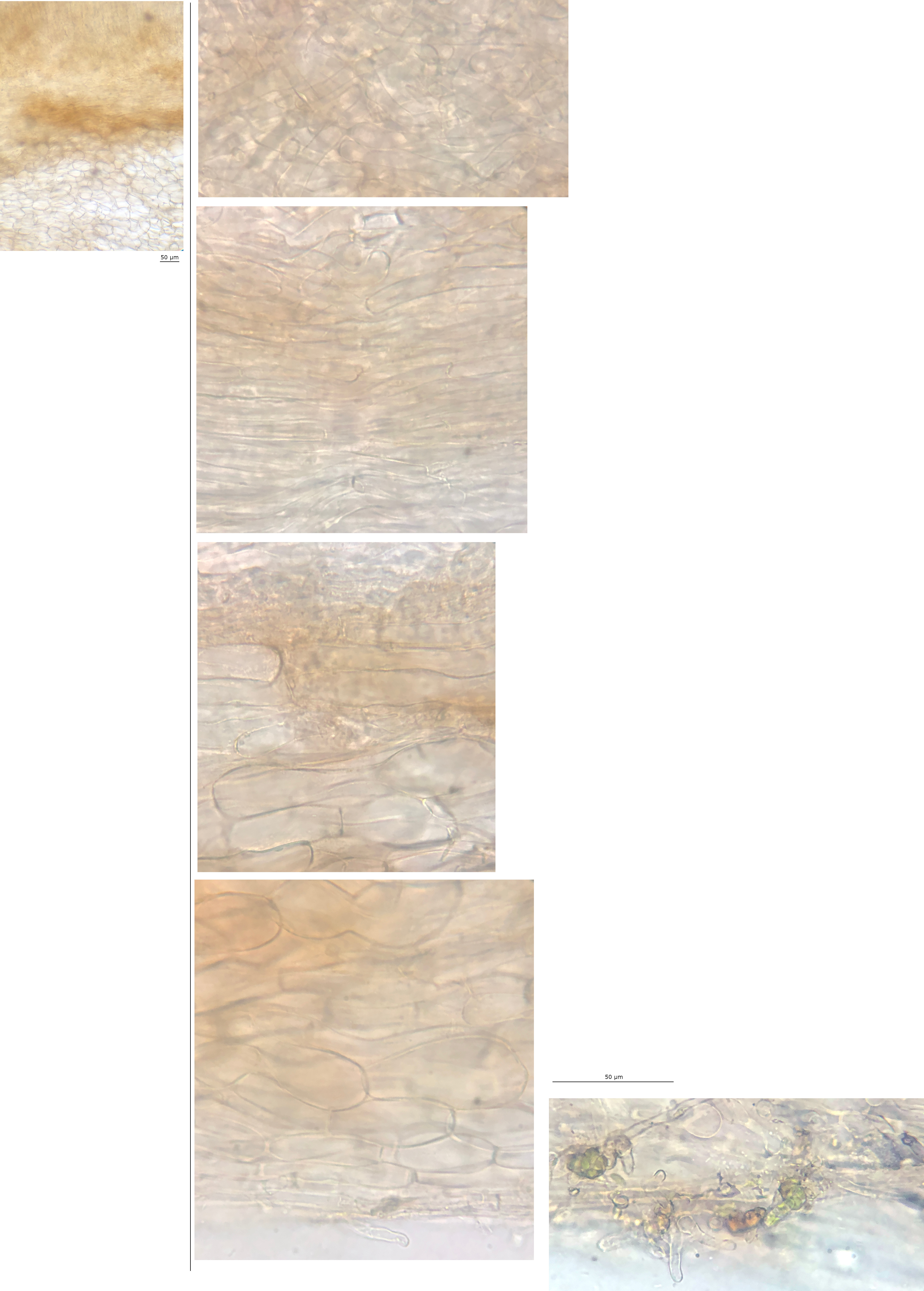 Excipulum-0003.jpeg