
20-05-2026 21:49
Margot en Geert VullingsWe found this Lachnum on Juncus stems mown last ye
20-05-2026 12:57
 Ingo Ibelshäuser
Hello everybody, on decayed hardwood e.g. Quercus
20-05-2026 20:08
 Andreas Millinger
Good evening,another quite distinctive find from M
20-05-2026 17:47
Margot en Geert VullingsWe found this Mollisia on dead Juncus stems mown l
22-04-2026 20:54
 Enrique Rubio
Hi to everybody.This Pyrenopeziza grew in moist le

19-05-2026 12:55
 Hardware Tony
After checking Gminder and Otto's library I cannot
19-05-2026 10:27
Patrice TANCHAUDBonjour, récolte récente sur terre retournée i
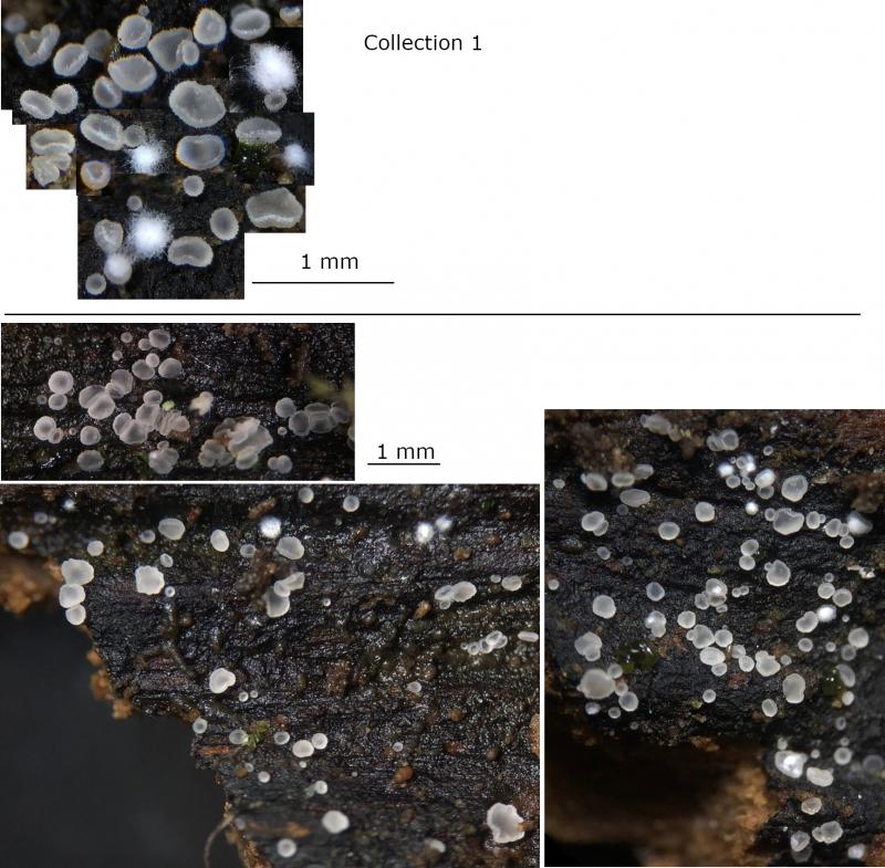
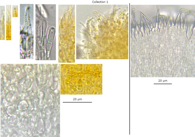
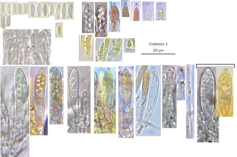
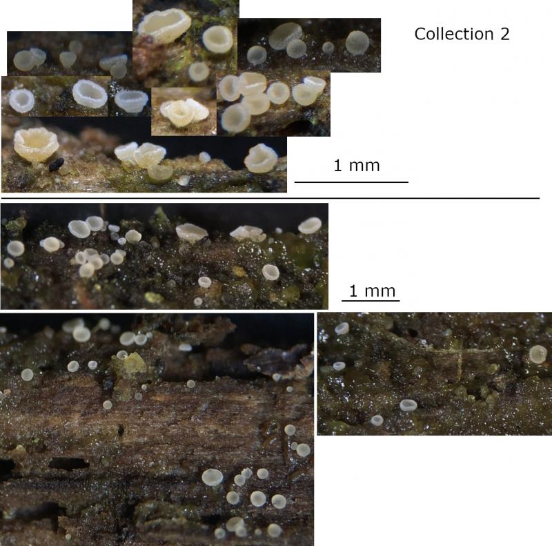
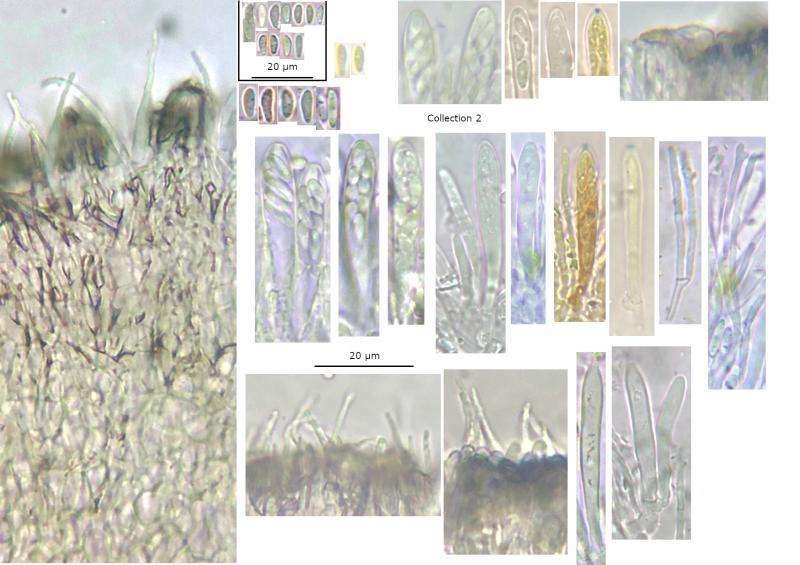
 • Hyaloscyphaceae (no VBs), Hyaloscypha: Macro and habitat, confirmed by spores and hairs (and paraphyses).
• H. fuckelii: Croziers, IKI bb and dextrinoid contents in hairs and paraphyses, spore size and ellipsoid-cylindrical shape, narrowly conical to lageniform hairs with thin-walls and light encrustation in water, some hairs septate or with apical knobs, some hyaline exudate, on +/- bulky deciduous wood.
• Frequent appearance in March seems to fit with Huhtinen's description of March-June as the peak season.
• In all three recent collections, some spores are longer or wider than Huhtinen's ranges for fresh in water (n = 30), although for CB + MLZ are more fitting (n = 604). The spores are also slightly wider on average compared to Huhtinen's measurements.
• Some apothecia in first observation overtaken by +/- localised white fluffy mycelium.
• Last observation on the ground often has less noticable hairs in macro, and more yellowish exudate in macro and micro.
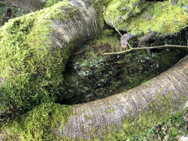
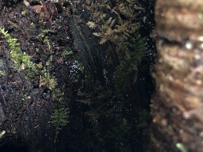
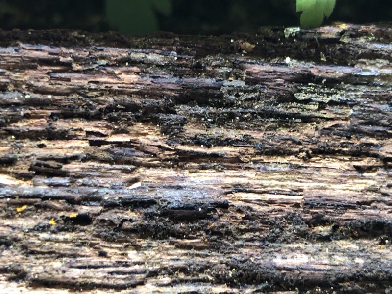
Habitat: Two observations within 100 m, on decorticated deciduous wood, no bodies of water in the immediate vicinity, in mixed deciduous woodland, southern England, mid-March, after generally wet weather.
• Observation 1: Inside a tree hollow, on brittle and blackened wood, sheltered under the overhang, ~2 m from the ground, in the trunk of a large Betula pendula, tree living and standing, damp when observed but possibly xeric.
• Observation 2: On a log on the floor, ~20 cm diameter, often on thick algae/biofilm, unidentified deciduous wood, with heavily decayed and pitted surface, appears hygric.
Associates:
• Obs 1: Dematiaceous fungi, algae, bryophytes.
• Obs 2: Algae/biofilm.
Storage and methods: +/- half sections from a single mature-looking apothecium in each collection examined, mounted in tap water, some squashing to separate cells, IKI or KOH added to the same mount.
Measurements of vital spores in water mount or asci:
• Collection 1: (5.8) 6.1 - 8.6 (9.9) × (2) 2.2 - 2.7 (3.3) µm, Q = (2.3) 2.5 - 3.7 (4.9), N = 41, Me = 7.3 × 2.4 µm, Qe = 3.
• Collection 2: (5.5) 6.2 - 7.8 (8.9) × (2.1) 2.2 - 2.7 (2.9) µm, Q = (2.1) 2.5 - 3.2 (3.7), N = 26, Me = 7 × 2.5 µm, Qe = 2.8.

I think I need to be more careful about how I measure small spores. The estimates were ok for identification but not more detailed comparison. In future I will be more careful about measuring small spores unless I have a very clear and flat view with oil immersion.
I have reviewed the measurements more carefully, and most spores are indeed smaller than 9 x 2.5 µm, but a small number (< 5%) do seem to measure slightly longer.
Current collection 1: (6) 6.2 - 9.1 (9.3) × 2 - 2.4 (2.5) µm, Q = (2.6) 2.9 - 3.8 (4.2), N = 20, Me = 7.3 × 2.2 µm, Qe = 3.3.
Current collection 2: (5.6) 5.9 - 8.6 (8.9) × (1.9) 1.91 - 2.2 (2.3) µm, Q = (2.8) 2.9 - 4 (4.1), N = 14, Me = 7 × 2.1 µm, Qe = 3.3.
Previous in vitro collection: (6.4) 7 - 8.9 (9.9) × (1.9) 2.2 - 2.48 (2.5) µm, Q = (2.8) 3 - 3.8 (4.5), N = 36, Me = 7.9 × 2.3 µm, Qe = 3.4.
I use piximetre to measure from photos. In some ways it helps to accurately measure the lines and get perpendicular axes, but it is reliant on the photo resolving the correct part of the spore as you mention.
Those results are copied directly from piximetre, but I would usually report to one decimal place as it does seem impossible to be sure about further distinction under the microscope.
I guess distinguishing between 2.4 and 2.5 um at 1000x is essentially distinguishing between 2.4 and 2.5 mm, which is certainly hard even at close reading distance. It is easier at higher levels of magnification but then the resolution of the microscope image is not sufficient.