23-02-2026 11:22
Thomas Læssøehttps://svampe.databasen.org/observations/10584971

29-11-2024 21:47
Yanick BOULANGERBonjourJ'avais un deuxième échantillon moins mat

07-02-2023 22:28
Ethan CrensonHello friends, On Sunday, in the southern part of

19-02-2026 17:49
Salvador Emilio JoseHola buenas tardes!! Necesito ayuda para la ident

19-02-2026 13:50
Margot en Geert VullingsWe found this collection on deciduous wood on 7-2-

16-02-2026 21:25
 Andreas Millinger
Good evening,failed to find an idea for this fungu
Possible Hyaloscypha albohyalina
B Shelbourne,
15-03-2024 13:30
• Hyaloscypha: Macro, no subiculum, tapered white (hyaline) hairs, spores, IKI bb, substrate.
• Seems to fit H. albohyalina: Croziers, IKI bb, spore size and shape, hair shape and ornamentation, traces of hyaline resin, ascus size, substrate.
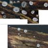
Habitat: On a sample of wood from a fence rail, ~5 x 2 x 0.5 cm, unidentified deciduous wood, collected close to a pond in mixed deciduous woodland in southern England in January, developed in vitro after almost 7 weeks, stored in a vented, transparent, plastic box with a damp paper towel, kept in the shade at room temperature.
Associates: Mollisia lividofusca that is still producing a small number of apothecia, originally some M. lividofusca apothecia in the population were infected with a 'chytrid' (and/)or a Helicogonium sp., algae still alive.
Preparation and methods: A central and an edge section from a single large and mature apothecium examined, mounted in tap water, no (intentional) squashing, IKI added to the same mount later.
Apothecia: ~15-20, scattered to gregarious, one cluster of 2, mixed maturity, whitish-greyish, superficial, sessile, initially globose and cupulate, receptacle apparently opening at a young age, becoming more broadly cupulate, +/- thin, broadly to narrowly appressed, loosely attached with no subiculum, texture soft-gelatinous; hairs short, white, straight, extending above disc, plentiful around margin and flanks, sometimes more sparse on flanks, developing early; margin usually distinct, slightly raised, more whitish and opaque than disc; disc shallow, becoming plano-convex, translucent, greyish in centre, frosted-gelatinous appearance.
IKI: Rings bb with calycina-shape, SCBs? in hairs and paraphyses dextrinoid (orange-brown), clumps of exudate yellowish, some paraphysis cytoplasm and spores yellow.
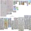
Asci: Cylindrical-clavate, croziers, 1-2-seriate with one spore at the apex; discharge by poricidal opening, happening in water mount, sometimes little to no (amyloid) ring material remaining, rarely appears torn (could be artificial), moderately to slightly turgid afterwards.
Vital mature: ~45-60 x 6-7 µm, apex rounded-conical, no apical thickening, 2-seriate, obliquely arranged, pars sporifera ~30-40%.
Dead: ~40-50 x 4-6 µm, apex more acute-obtuse, more cylindrical, occasionally very narrow apex giving lageniform shape, apical thickening ~0.75-1.25 µm, 1-2-seriate, more vertically arranged, pars sporifera ~50-80%.
Spores: Allantoid-cylindrical to ellipsoid, occasionally fusiform, inequilateral and usually curved in profile view (reniform), poles rounded-acute, usually slightly heteropolar with the base more attenuated and acute, several small and shadowy LBs towards each pole, LB OCI 1-2, a larger clear LB towards each pole, no septation observed.
Measurements of vital mature spores discharged in water mount:
(6.4) 7 - 9 (10.1) × (1.9) 2.2 - 2.6 (2.7) µm, Q = (2.4) 2.7 - 3.8 (5), N = 40, Me = 7.9 × 2.4 µm, Qe = 3.3.
Paraphyses: +/- cylindrical, apex rounded, 2(-3?)-septate, apical cell often longer ~1.5-2x, occasionally a medium-size dextrinoid SCB? in some cells (difficult to see in section in water), dichotomous branching occasionally observed at basal septa, not extending above asci.
Exudate: Minimal in water mount and creating oily residue, hyaline, resinous, occasionally clinging to hairs in clumps.
Hairs: ~35-50 x 2.5-4 µm, apices ~0.5-1.25 µm wide, narrowly conical-attenuated, straight to bent, occasionally very slightly sinuous, generally aseptate but occasionally uniseptate below, hyaline, rough appearance due to tiny warts, occasionally with a hyaline resinous lump (remaining in IKI), often hyaline-yellowish and dextrinoid SCB? at apex, no branching; warts scattered, mostly tiny and shadowy, occasionally slightly larger and greenish, usually more prominent around apex, remaining in IKI.
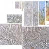
Ectal excipulum: Hyaline, textura prismatica (imbuta), thick walled, cylindrical-clavate marginal cells (without hairs) dispersed amongst hairs; SCBs?, occasionally one in a cell, medium to large, globose, greenish.
Medullary excipulum: Clearly separated by horizontal hyphae with thinner walls, hyaline, textura intricata.
Subhymenium: Hyaline, many swollen-globose cells, occasional SCBs like ectal exipulum, often a tiny shadowy LB in each cell.
Basal attachment: Some hyaline anchoring hyphae, not forming subiculum.
Hans-Otto Baral,
15-03-2024 15:30

Re : Possible Hyaloscypha albohyalina
H.albohyalina has larger spores. H. fuckelii or H. vitreola?
B Shelbourne,
15-03-2024 17:25
Re : Possible Hyaloscypha albohyalina
Thank you, I'm not sure why I missed that as the range and average are higher in Huhtinen's monograph. It must be a statement in the key that I misinterpreted. The spores are too small for H. vitreola too, it must be H. fuckelii.
Some hairs have an apical knob, and looking again there is some septation in the upper part, which are both characteristic of H. fuckelii. I thought the warts were quite conspicious but I haven't seen any other species to compare it to.
Some hairs have an apical knob, and looking again there is some septation in the upper part, which are both characteristic of H. fuckelii. I thought the warts were quite conspicious but I haven't seen any other species to compare it to.


 Hymenium-0004.jpeg