

05-05-2026 22:40
Gernot FriebesHi,I believe this is a Plagiostoma growing on a Sa
06-05-2026 11:25
 Castillo Joseba
Me mandan el material seco de Galicia (España) re
06-05-2026 17:23
Thomas Læssøehttps://svampe.databasen.org/observations/10594257
28-04-2026 20:07
 Lothar Krieglsteiner
... on twig in the air at standing Ceratonia siliq
04-05-2026 18:13
 Stephen Martin Mifsud
ID request for what seems to be a true aquatic fun
04-05-2026 16:39
Stephen Martin Mifsud
ID request: This specimen was collected in Malta o
28-07-2011 18:31
 Alex Akulov
Dear FriendsToday I made the pdf file of Velenovsk
04-05-2026 09:50
Castillo Joseba
Me mandan el material seco de Galicia,(España) re
 Discomycete with Stilbella-like anamorph, seems to have similarities with genus Claussenomyces (s.l.). Possibly in the C. prasinulus group (Dendrostilbella) as no ascoconidia, and possibly differing from C. prasinulus in the colour of the apothecia, lack of subiculum, and some characters of ascospores. I noticed some similar looking observations on the forum too, maybe the 'waldhaff' species.
Files also included for some photos as may not loose resolution in upload.
I have tried to use the monograph of Seifert (1985) to help with assessing morphology of the anamorph and couldn't find a modern or comprehensive treatment of the genus or group.
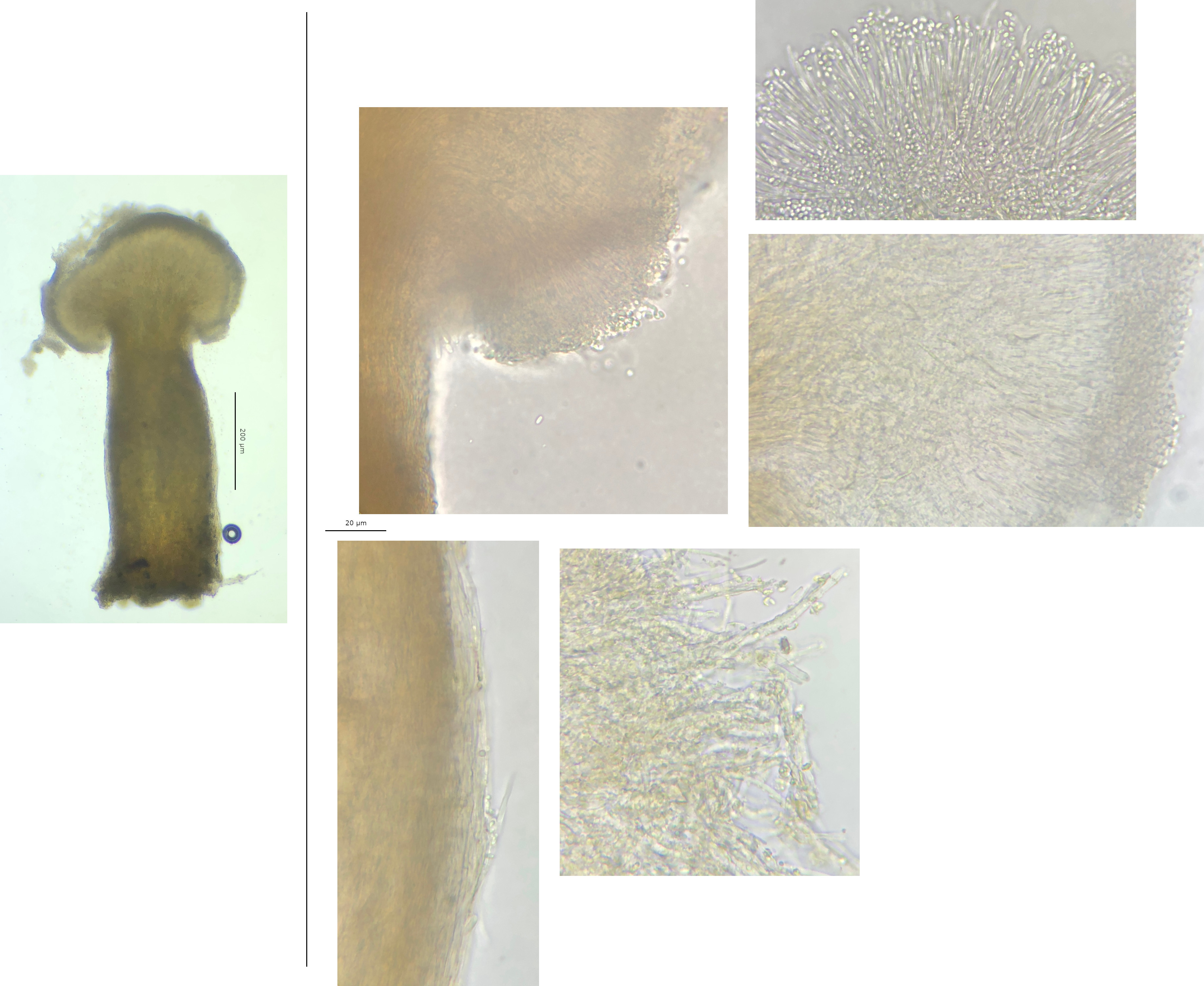
Preparation and methods: Stored in damp box attached to a piece of wood for a day or so. A single synnema and two central sections of an apothecium were examined, mounted in tap water, and later lightly squashed. IKI (~1%) applied to squashed apothecium sections after some time.
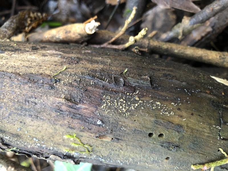
Habitat: On unattached decaying branch, hygric and saturated at the time, decorticated part, from unidentified deciduous tree, lying close to the ground, in a loose pile of sticks and small branches, damp and shady area, in mixed deciduous woodland. Found a few days ago (February) in southern England.
Associates: Mollisia sp, dematiaceous fungi, very young lichen, algae, bryophytes, a large mite (seemed to like eating the apothecia of the Mollisia sp).
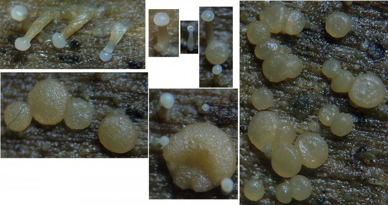
Sporomata: Stilbella-like synnemata broadly distributed, discoid apothecia in several groups, often 1 or 2 synnemata basally attached or closely associated to an apothecium, apothecia seem to arise later.
Anamorph
Synnemata: Scattered to gregarious, rarely clustered or branching basally into two, parallel determinate (parallel stipe hyphae and capitate), robust to medium stature (Q ~3).
Stipe:
• Macro - subulate to cylindrical, occasionally bulbous, initially whitish transluscent, then straw-yellowish going browner, fibrous appearance, quite broad.
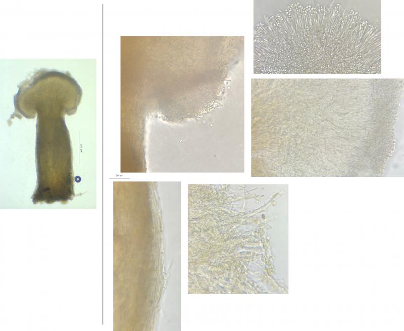
• Stipe hyphae - parallel, textura porecta, marginal hyphae with several small LBs and unornamented, marginal hyphae at apex, yellowish-orange exudate, exudate remaining in large clumps in water and attached to hyphae after squash.
Capitulum:
• Macro - creamy off-whitish, gelatinous appearance, initially globose becoming more flattened and pulvinate, some with brownish patches, fusing when touching, powdery residue remaining when overmature.
• Micro - formed of dense palisade of phialidic conidiophores, arising from stipe hyphae, covered by a large mass of conidia ~20 – 30 µm thick, no exudate visible but conidia adhering to each other.
Conidiophores: 1 – 2 verticiliate, sometimes irregular, stipe-part often with several small but distinct warts, warts protruding ~0.75 µm.
Metulae: In whorls of 1 – 4, ~17 – 20 x 1.3 – 1.5 µm, some small to medium size LBs.
Phialides: In whorls of (2) 3 – 6, ~11 – 14 x 0.75 – 1.5 µm, narrow lageniform (tapering slightly towards collarette and collarette not flaring), narrowly spatulate with conidium attached.
Conidia: Aseptate, unornamented, cylindrical to ovoid, yellowish en masse, adhering to each other (sticky), OCI 2 – 3, 1 – 2 medium-size VBs often near the poles, VBs green and diameters ~0.5 – 0.7 µm.
Conidia with synnemata:
(2.3) 2.5 - 2.9 (3.1) × (1.3) 1.4 - 1.7 (1.9) µm,
Q = (1.5) 1.6 - 1.9 (2), N = 30,
Me = 2.7 × 1.5 µm, Qe = 1.8.
Conidia on apothecia (almost identical measurements):
(2.2) 2.4 - 2.9 (3.1) × (1.1) 1.3 - 1.7 (1.9) µm,
Q = (1.3) 1.5 - 2 (2.5), N = 30,
Me = 2.6 × 1.5 µm, Qe = 1.8.
Subiculum: None or very limited.
Teleomorph
Apothecia: Gregarious to caespitose in small groups, translucent straw-yellowish, getting darker, initially urceolate to turbinate, becoming more pulvinate, occasionally cupulate due to local pressure, substipitate, waxy-gelatinous appearance, consistency relatively firm, margin unpronounced and mostly even, disc almost indistinguishable but slightly lighter coloured, disc plane to convex and occasionally uneven/lumpy. Strong similarities to colour and texture of the stipe of synnemata (related to exudate?).
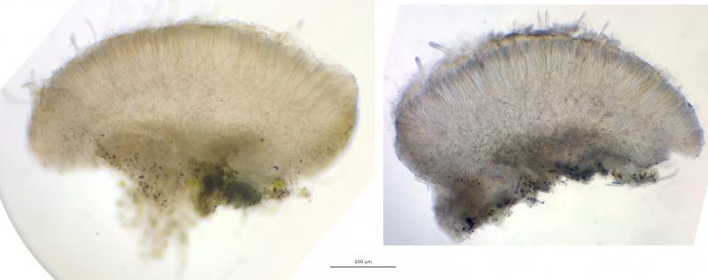
Epithecium: Orangish, ~70 µm thick, ~50% extending into hymenium, mature asci protruding, paraphyses protruding?, some piles of sticky conidia (possibly transferred from synnemata), remaining in large chunks in water after squashing.
Asci: Rounded apex, croziers +(/-?), thin-walled, possibly poricidal opening, often with swelling above base, no inversely oriented spores rings and wall IKI -, ascoplasm dextrinoid, some contents going strongly red (particularly around apex).
- Mature vital (turgid): Mildly clavate, apex rounded, no sign of apical thickening, spores angled and tight packed in apex, 2 – 3 seriate and overlapping, spores often in symmetric pairs, pars sporifera ~20%, 155 – 192 x 11 - 14 µm.
- Mature dead (presumed, flaccid): Cylindrical, large apical thickening 1 – 6 µm, sometimes with +/- acute apical canal in front view ~1- 2 µm tall, rarely small apical pendants visible, spores vertically oriented, 1 – 2 seriate, pars sporifera >40%, 100 - 160 x 8 - 11 µm.
Ascospores: Croissant-like appearance, allantoid-fusiform, lunate in profile view (curved), poles rounded to acute, often heteropolar, then upper pole wider and more rounded with lower pole tapering to acute, (3) 6 – 7 (9) septa, mostly horizontal septa but rarely vertical too, thick walled?, often narrowing at septa, rarely appearing to fracture or strongly narrow around a central septum, no budding (rarely from either end?), IKI yellow and VBs less visible.
- Vital: Each cell with 1 – 4 (6) medium to small VBs and several LBs, VBs appear to be shared into cells, OCI 4 – 5+, VBs greenish with diameter ~ 1 – 2 µm.
Vital spores discharged from section in water mount:
(14.3) 15.1 - 20.5 (22.9) × (3.7) 4.2 - 4.9 (5) µm,
Q = (2.9) 3.4 - 4.7 (5.3), N = 30,
Me = 18.4 × 4.5 µm, Qe = 4.1.
- Dead (presumed): No visible contents, cells more flaccid, and septa clearly visible.
Paraphyses: Narrow-filiform, sinuous, multi-septate, apical cell often slightly longer, occasionally branching - even close to the apex, several smallish VBs, VBs hyaline, occasional small shadowy LBs, appear to extend above asci.
Medullary: Dense hyphae, textura intrica, hyaline.
Ectal: Textura globosa, hyaline, brownish at base, thin-walled, marginal cells elongated, exudate like epithecium.
Hairs: Possibly short and hypha-like.
Subiculum: None seen in macro, a few brown hyphae observed around base but may be unrelated.


 Lower-details-0001.jpeg
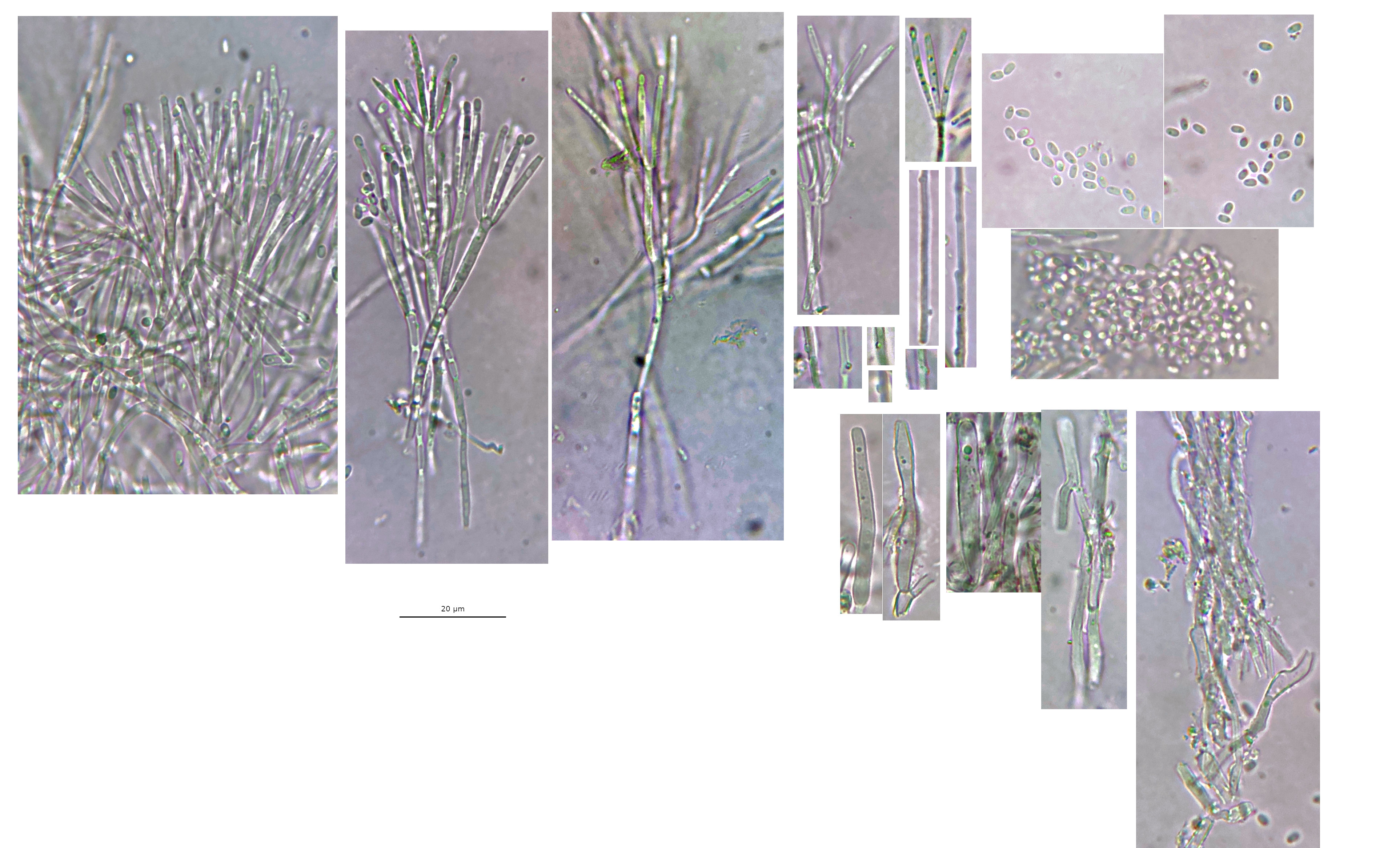 100x-details-0001.jpeg
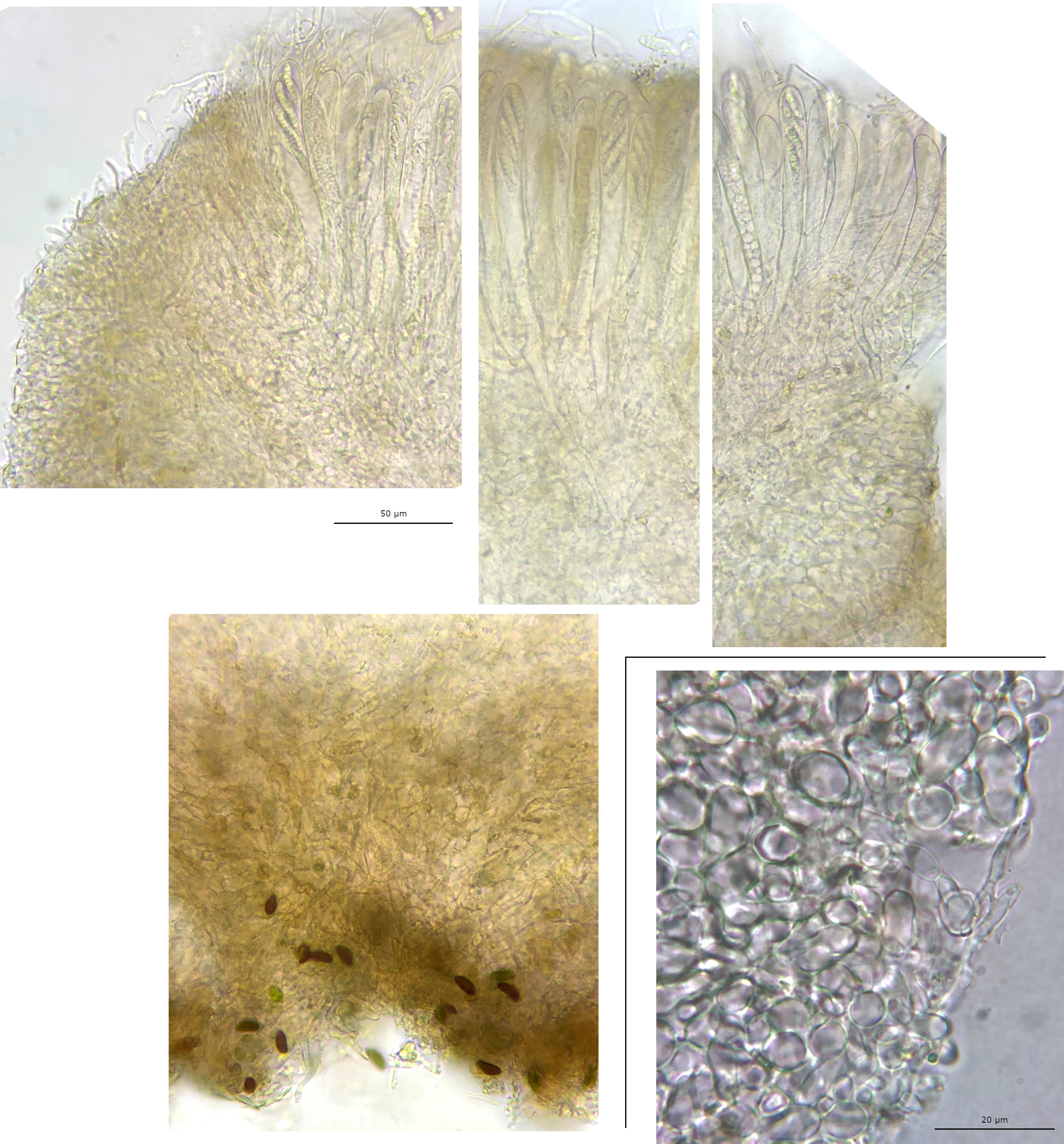 40x-details-0001.jpeg
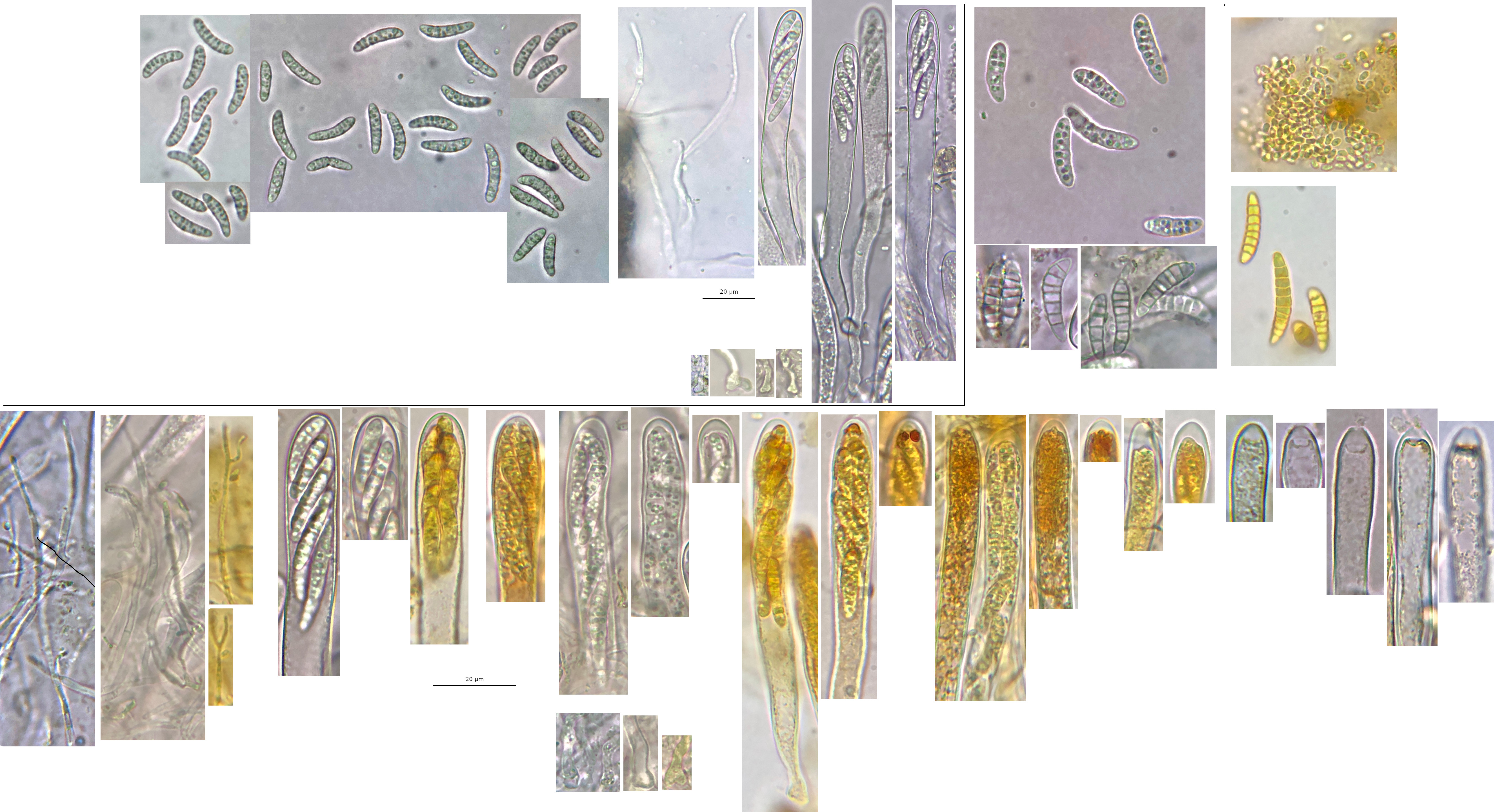 Hymenium-details-0001.jpeg
Seifert (1985) reports the teleomorph of C. prasinulus to be often with an inconspicious whitish subiculum, possibly following Dennis (1956). Looking at photos of C. prasinulus online, including your folder, occasionally there seems to be a localised whitish subiculum surrounding an apothecium or a synnema. The subiculum may just be unusual (and not present in my collection) rather than differentiating.