11-05-2026 12:32
 Bernard CLESSE
Pourriez-vous m'aider à identifier cette héloti
13-05-2026 15:26
 François Freléchoux
Bonjour,Voici une récolte faite il y a quelques j
12-05-2026 15:41
 Nicolas VAN VOOREN
Dear Ascolovers, especially interested in Pezizale
13-05-2026 12:05
 Thierry Blondelle
Bonjour à tous,J'aimerais avoir confirmation de c
10-05-2026 23:17
 Andreas Gminder
Hello,today we found in a moist steep decidous for
28-04-2026 20:07
 Lothar Krieglsteiner
... on twig in the air at standing Ceratonia siliq
27-04-2026 20:52
Lothar Krieglsteiner
Found on hanging tiwg of Olea europaea in dried-ou
11-05-2026 20:22
Lothar Krieglsteiner
on attached twig of standing Ficus caricaquite uns
29-04-2026 10:44
Lothar Krieglsteiner
growing at moist, drying-out soil at the side of a
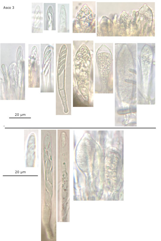
 Studying three ascomata from a suspected Mollisia sp., I found that two had a mixture of 'normal' and very swollen, opaque asci. It is difficult to speculate on the cause without more context, and I would appreciate any suggestions.
It may be a relatively simple answer like over-maturation, but the appearance is quite different to the other asci (even ones that look vital and turgid to me) and those in the few ascomata of other species I have studied previously.
The first ascoma examined was stored for several days prior (in a damp container), but the following two were collected later and examined within hours of collection. Except the missing swollen asci in the second ascomata examined, the characters are similar in the three ascomata, and my current working assumption would be that all are from the same species.
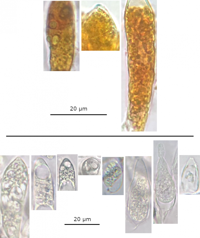
Inflated asci:
• Contents - a large, relatively opaque group of jumbled circular-looking contents, difficult to interpret these inner contents with any confidence (possibly spores and LBs), some with large LBs around base, sometimes opaque contents fills ascus or transparent areas at the base or apex.
• Shape - more broadly cylindrical to ellipsoid, and some look less turgid with the contents at the bottom giving an asymmetric lageniform (bottle-shaped) appearance (particularly in the stored ascomata).
• Apex – seems like 'normal', but the contrast causes a more or less pronounced mammiform appearance, also seems harder to see the IKI bb reaction.
• Base – again like 'normal', both with croziers.
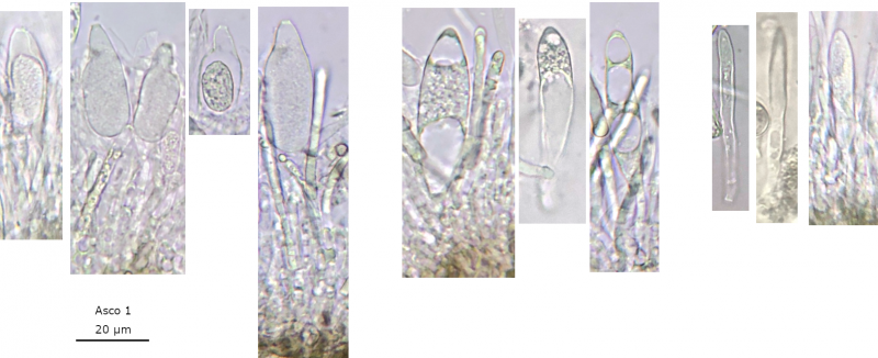
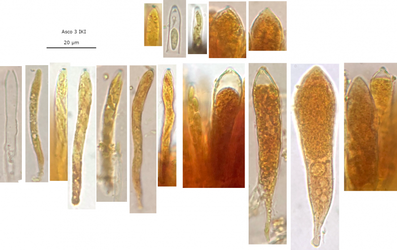
Your lugol treatment seems to show that you have some unparasitised asci of the host. (Narrow asci with amyloid (blue) reaction). The swollen asci are inamyloid (no blue reaction).
The parasite causes the host's asci to become swollen and the parasite's spores develop inside these swollen asci.
If you have a Pyrenopeziza instead of a Mollisia, then there is another Helicogonium species which will parasitise these. Therefore the Helicogonium species attack ascomycetes from a specific genus.
Another example is H. orbiliarum which attacks Orbilia fruit bodies.
With Best Wishes,
Peter.
This seems to be a good explanation. It does seem like the larger asci do not have an IKI reaction and the faint blue colour on some could just be a refraction on the tip of the apex. I also thought that I could see too many spores inside these asci and some of these looked quite different but I didnt want to prejudice things by speculating too much.

Okay. Since I found this parasite in Mollisia fruit bodies in July 2014, further work has been undertaken, such that Helicogonium is no longer considered correct.
I will have a look through your folder.
Does this also apply to Helicogonium orbiliarum and other similar invasive parasites?
Thank You,
With Best Wishes,
Peter.
From the illustration in the monograph I had determined at least some of these swollen 'asci' were sporangia from these 'chytrids'. Looking at your pictures in the folder and the illustrations of Helicogonium asci in the monograph, it does seem like there are only these sporangia.
The broad similarity of these sporangia to asci is quite interesting to me, but I guess both are rather primitive. The mammiform apex in particular seems distinguishing from asci of Helicogonium, and I guess studying the base more careful and the plasmatic contents.
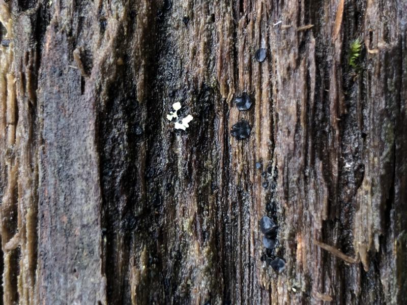
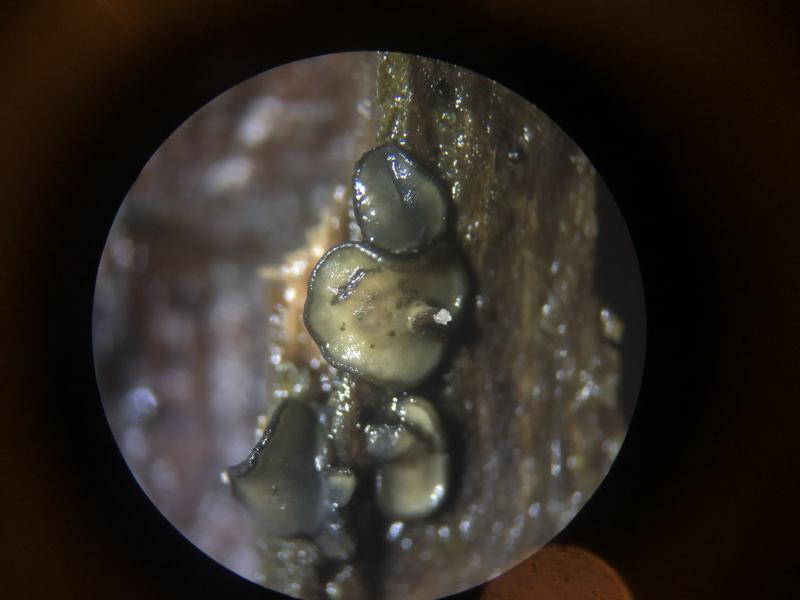
Another similarity I noticed with some of the photos in your folder is a mixture of ascomata with darker and lighter grey discs, I wonder if this is related to the parasitisation of the ascomata or just a character of a species that is parasitised. I have attached a photo for reference.
I will have a go at identifying the Mollisia sp. now that I can hopefully distinguish, and I can review my microphotos to determine what else be related to this chytrid, and not an algae or something else that I may have assumed when looking.
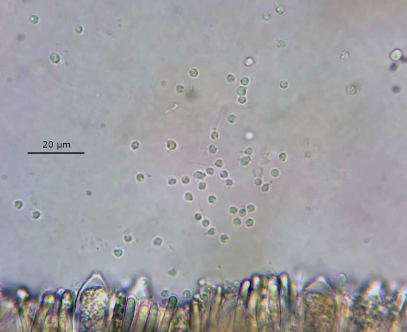
In hindsight, it seems that in some of the sporangia I saw there are a (significant) number of globose to ovoid (zoo)spores, each appearing to have several VBs. This can almost be seen in some of the better resolved microphotos I posted previously, and the collage attached.
I have also been thinking about a group of similar looking objects, found in a group near the hymenium of a section taken from the third ascomata within hours and examined in tap water. These proceeded to become increasingly motile, which was annoying at the time as I was trying to view and photo spores of the host. Looking at some videos of chytrid zoospores, the broad morphology and pattern of movement seems similar.
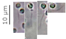
I have also attached the only good microphoto I took of these suspected zoospores. It was very challenging to resolve any flagella, but I also attach some extracts from this the photo that appear to show chytrid-like (singular, posterior, whiplash) flagella, as suggested by the movement. Some observations could suggest intra-sporangial release.