
17-04-2026 19:16
 Enrique Rubio
Hi to everybodyI would appreciate any assistance r
14-04-2026 05:32
Ethan CrensonHi all, A few weeks back a friend pointed out som
17-04-2026 15:14
 Bruno Coué
Bonjour.Récoltes du 16/04/2026, sur feuilles mort
12-04-2026 15:52
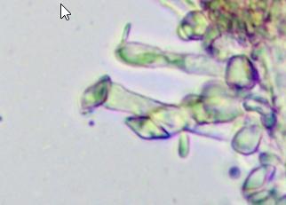
Gernot FriebesHi,I'm looking for help with this anamorph collect
14-04-2026 21:52
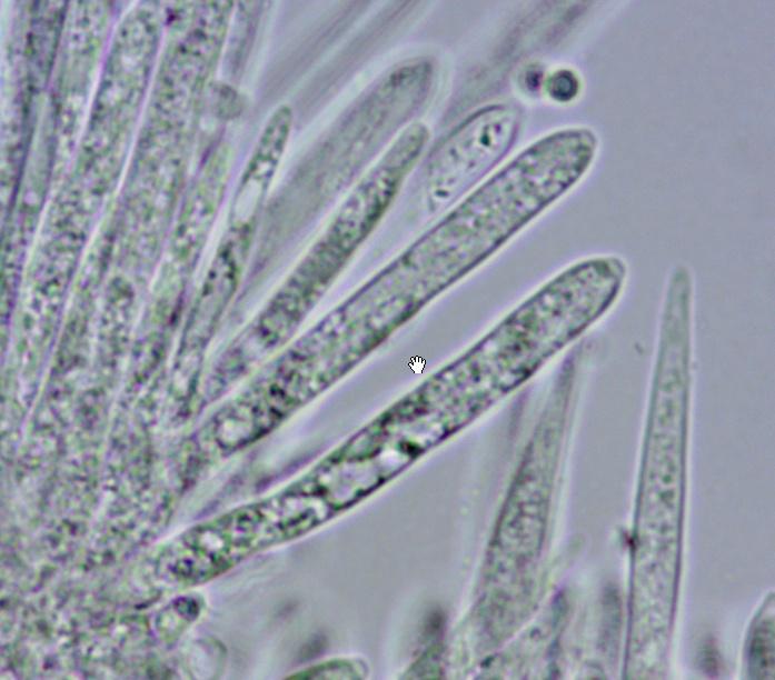
Gernot FriebesHi,found on dead leaves of Carex elata. Conidia: 4
16-04-2026 22:09
Buckwheat PeteHello, I'd like to ask about this older specimen:
15-04-2026 19:33
Fátima Durán ManzanequeHi!! I need help, I found this Ascomycete but I d
14-04-2026 20:31
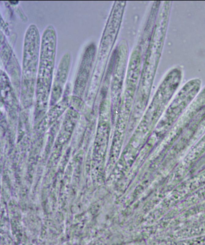
Gernot FriebesHi,can this be Psilachnum lateritioalbum on Phragm
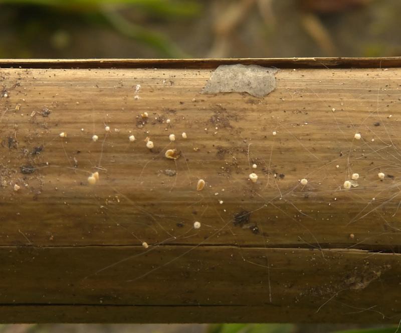
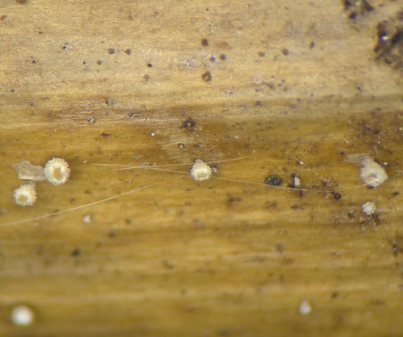
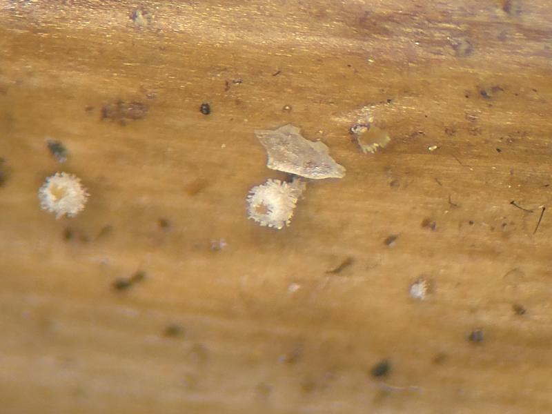
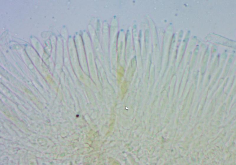
12-04-2026 17:56
 Hardware Tony
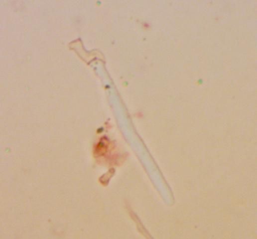
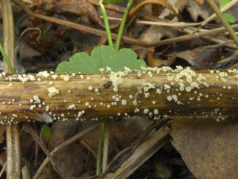
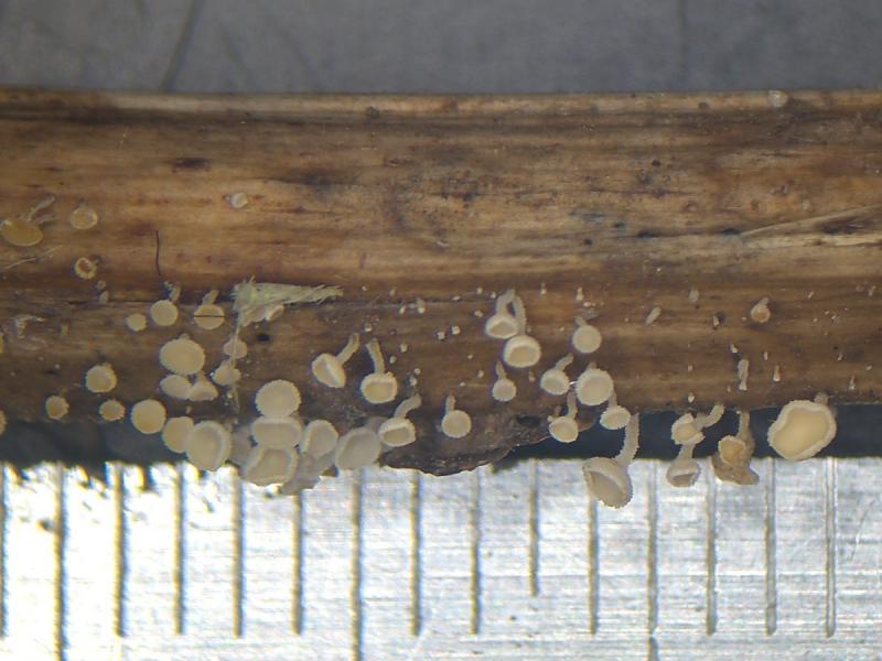
Found on dead stems in February earlier this year
12-04-2026 12:22
 William Slosse
In a dune grassland in Oostduinkerke (Belgium), on
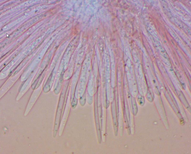

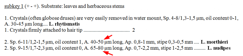
In my opinion is your find Lachnum nudipes.
Why do you think that the asci too small for L. nudipes?
Greetings
Ingo
One source is no source.
Look here on Zotto's IVV:
https://drive.google.com/drive/u/0/mobile/folders/0B5SeyOEkxxZhejlueDdoM3FqQkk/0B5SeyOEkxxZhdzZqNFRWZXBScHc/0B5SeyOEkxxZhUFlCTXZLYlk5eUE/0B5SeyOEkxxZhbXUwamduSmpaNGs?usp=sharing_eil_m&ts=629667c4&resourcekey=0-U2ZEbJo27Q5uPFiOVJHLyw&sort=13&direction=a
Greetings
Ingo

>>> I assume you compared your dead (shrunken) asci with my living asci.
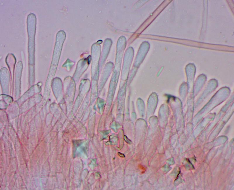
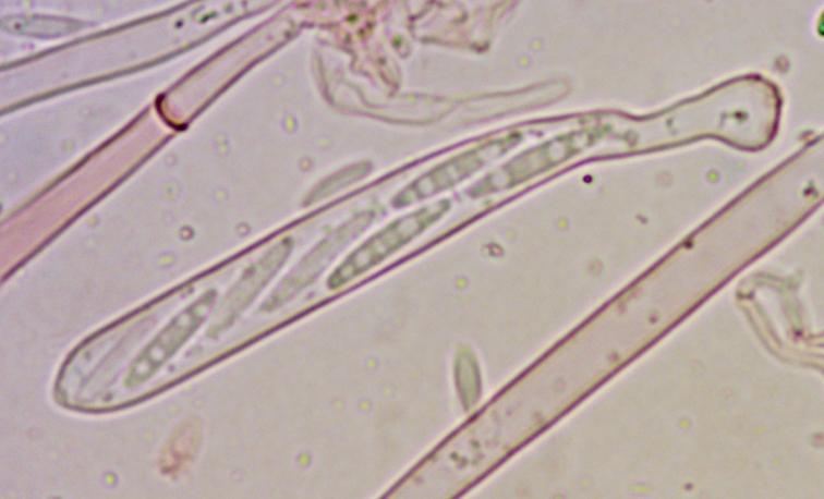
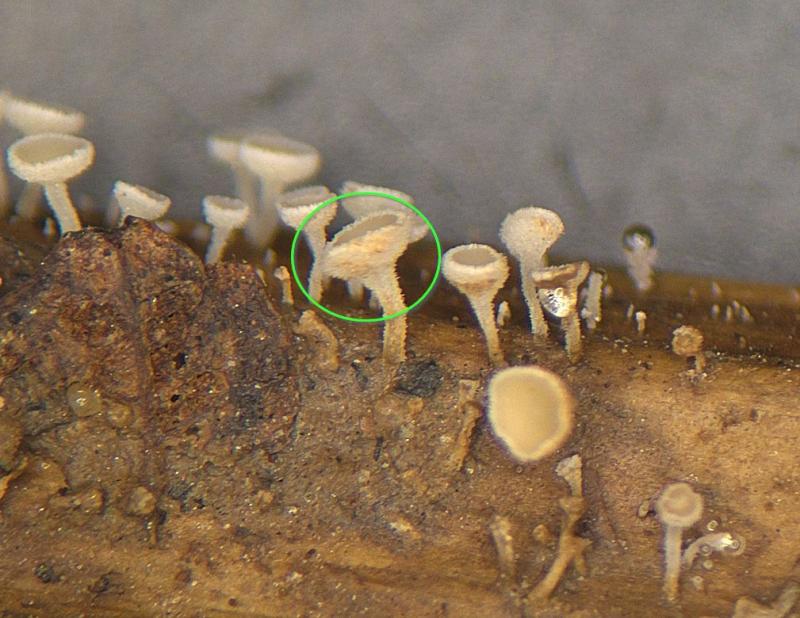
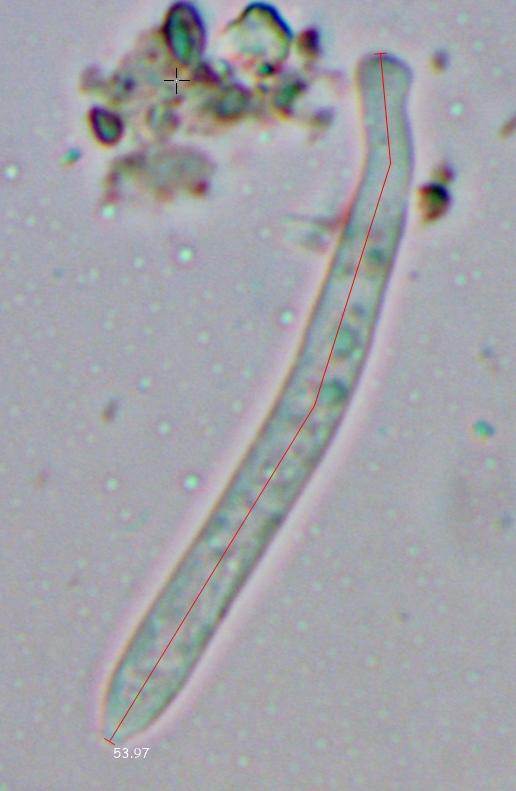
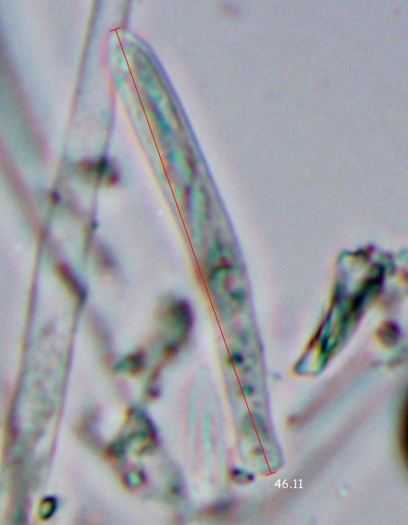
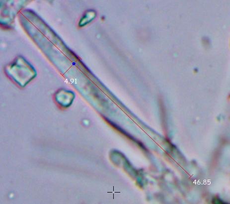
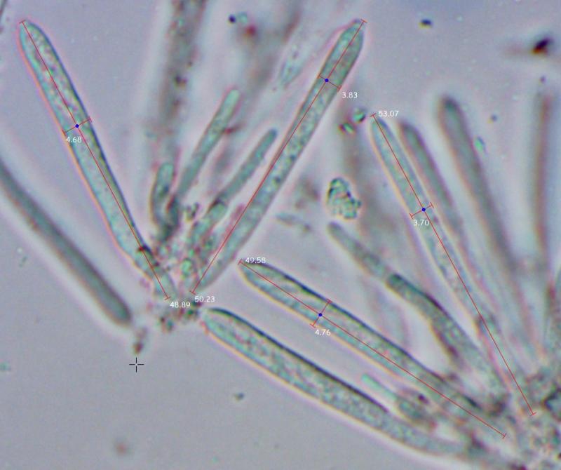
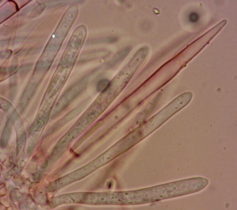
There were guaranteed living asci. I mounted the squashed part of the living frb immediately after collection, no more than 10 minutes passed from taking a sample from the nature with part of Filipendula stem, and no more than 10-20 seconds after removing frb from the substrate. Asci and spores were mounted in SDS/Congo red.
At this time part of the stem with these specimens lives in the humid container. I now (some minutes ago) examined one new specimen (marked on the attached photo) in the water mount. Asci was observed with 60x lens, ascospores - with 100x oil immersion lens. It was 100% living and fresh specimen. This study shows that previous measurements were correct.
asci - (46.1) 46.9 - 53.1 (54.9) × (3.4) 3.8 - 4.9 (5.3) µm
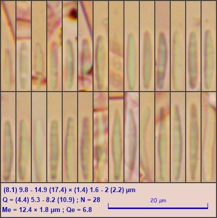
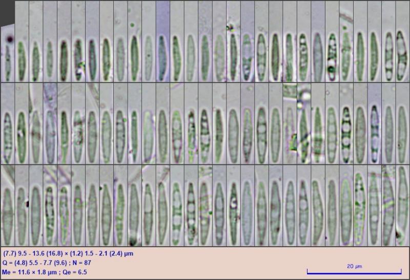
ascospores (now measured bigger set of spores):
(7.7) 9.5 - 13.6 (16.8) × (1.2) 1.5 - 2.1 (2.4) µm
Q = (4.8) 5.5 - 7.7 (9.6) ; N = 87
Me = 11.6 × 1.8 µm ; Qe = 6.5
About oily content - i see that some spores contain refractive content, some other spores contains non-refractive vacuoles, still others contain none of that. But maybe I'm misinterpreting what I see.
>>>>Did you ever look in my article in Mycotaxon, 1992, on vital taxonomy?
Yes, I often look at this article when I study ascomycetes under a microscope.
But in this case, all the requirements of the preparation were met. The material is fresh and not dehydrated (not even a little), there was no extensive squashing. In the last preparation no squashing at all was.
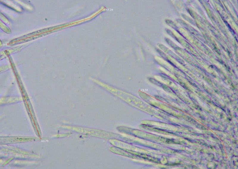
I thought that here the contents of the asci in the form of a large number of small and medium VB's, behind which the ascospores are almost not visible (new photo from the last mount with 100x immersion lens), indicates that the asci are alive (as in the paraphyses, as on fig.33 in the article mentioned). They should not have died, especially in the last careful mount.
For comparsion - microphoto #2 in Congo red/SDS of dead asci from the same frb - VB's in the asci are dissolved, and some asci clearly shows ascospores inside.
But it turns out that I'm wrong? In this case, I can't understand why asci are died in the last and pre-last mount and paraphyses not.
PS - previous photo was with 40x lens whos resolution can't clearly show small VB's as discrete objects.
>>> You used the term VBs for contents of the ascospores? Or asci? Or paraphyses?
I used the term "VB" in general for any content of vacuoles in all tissues, disregarding of their content type (refractive/non-refractive, lipidic, e.t.c.)
>>> Generally it is possible that your collection experienced some unfavourable events in the field. It can be that apothecia look externally sound but contain no living elements.
Highly likely this is my case. Temperature stress could be another cause of death besides death in the habitat. Being taken from a lying stem from under a layer of last year's leaves in a very wet place in dark dense thickets at a temperature of 12-14C (as surface groundwater), they were placed in a container with wet cotton, and transported for 10-20 minutes at a temperature of 22-24 C (like air). Usually samples of such small fungi, being taken in a wet habitat, not removed from the substrate and placed into in a moist container, continue to mature and begin to sow spores. But in this case, the fruiting bodies really stopped growing, which indicates their death.
I almost always study microscopic features of only fresh living material, and very rarely exsicates. I considered these samples to be a priori alive, and for me this is the first such case for more than 10 years. For me, the study of dead material is unusual. That's why I'm confused.
In any case, thank You very much again for your attention, now everything is clear and I should remember this case.