04-05-2026 16:39
 Stephen Martin Mifsud
ID request: This specimen was collected in Malta o
04-05-2026 09:50
 Castillo Joseba
Me mandan el material seco de Galicia,(España) re
28-04-2026 20:07
 Lothar Krieglsteiner
... on twig in the air at standing Ceratonia siliq
02-05-2026 12:42
Alain BRISSARDBonjour à tousJeuidi 30 avril dernier on m'a remi
02-05-2026 13:06
Pauline. PennaBonjour Please can someone help me with this id
01-05-2026 22:45
 Thierry Blondelle
Bonjour à tous, Une récolte sur bouse séchée d
14-04-2026 05:32
Ethan CrensonHi all, A few weeks back a friend pointed out som
28-04-2026 20:33
Vitus SchäfftleinHello, I found Trochila ilicina on Ilex aquifoliu
Dear colleagues. First of all I wish everyone a better new 2022 and hope things get back to normal soon.I am studying a Necteriaceae, possibly a Cosmospora it was similar to this entry in the af db :
http://www.ascofrance.fr/search_recolte/2236
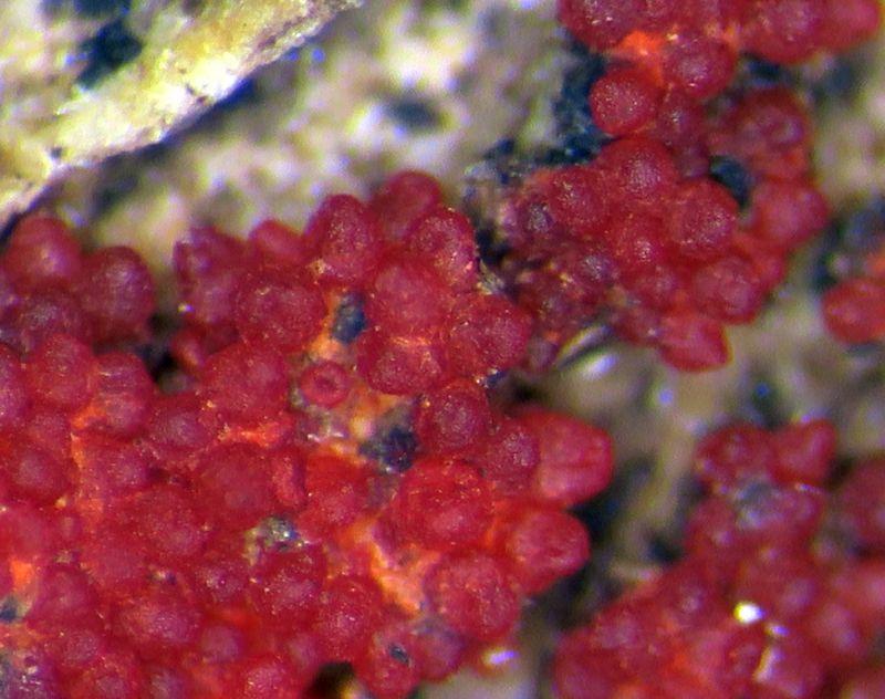
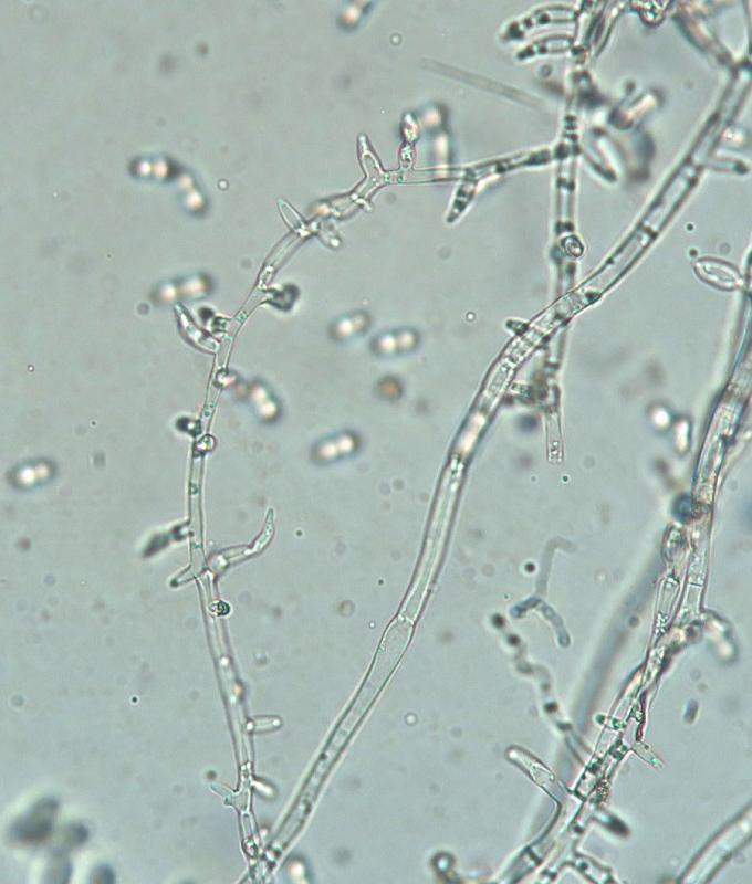
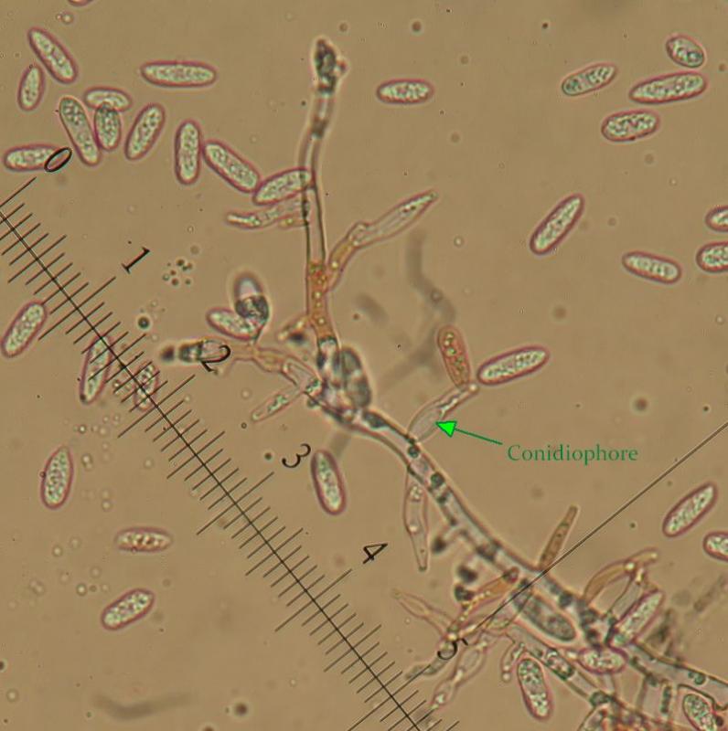
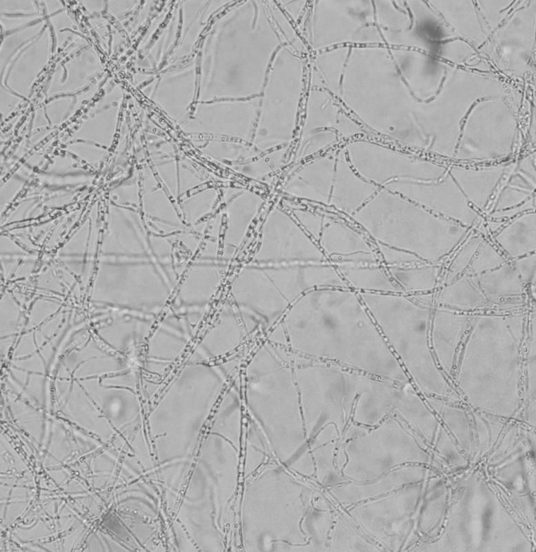
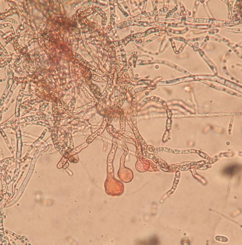
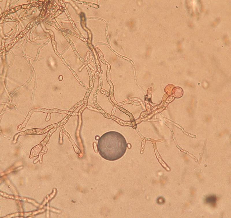
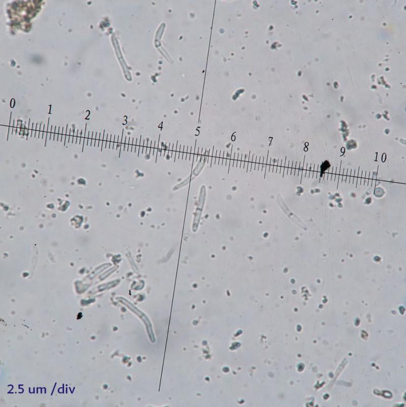
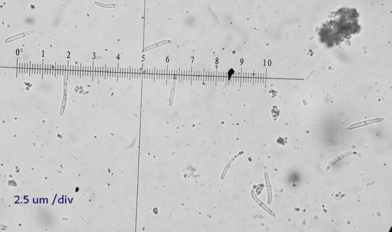
I have managed to isolate it as an anamorph but before further tests I want to confirm that the Anamorph I isolated do make sense. the mould produced those orange pustules which I saw in some site for cosmospora sp. so I think we are on the right track. The spores looks like that in the database labelled Cosmospora sp. by ALAIN GARDIENNET (link above). The hyphae have numerous oil bodies refractive bodies. I could not detect obvious conidiophores (maybe they just bud from simpile conidiophore-like branches. Spores 10-16 um long. It does not look like a fusarium :-(
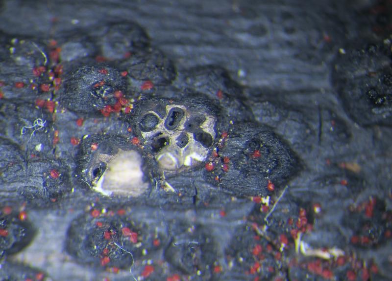
The Perithecia were growing on some black Xylariaceae stomata attached on fallen olive branches. They were pyriform in my opinion.
- - - - bottom line - - - -
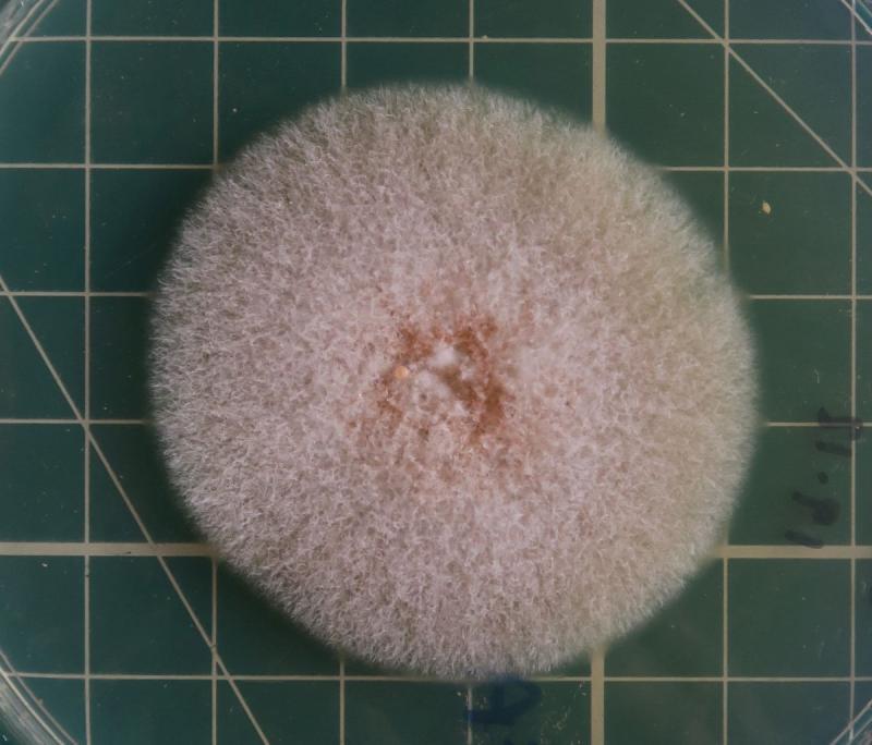
There was also some kind of contaminant fungus, so I subcultured on different media to try and have a pure culture. I would also be interested to know what is that 'contaminant fungus' too.
Thanks and hope this is interesting after lots of work! (I should take some more pics!)
There is a paper by Herera et al (2015) dealing with Pseudocosmospora....


Have you identified the host? This is very important characteristic in order to have a chance to know what your cosmospora-like fungus is.
Did you see asci and ascospores in your collection? It would be useful to make a vertical section of the ascomatal wall.
I have forgot to finish my sentence and I was saying There is a paper by Herrera et al (2013) dealing with Pseudocosmospora.... where the anamorphs have the same spores.
https://www.tandfonline.com/doi/abs/10.3852/12-395?journalCode=umyc20
My question was initially to be sure that I got the right anamorph isolated, maybe I progress to DNA seq. I know Cosmosporas should have Fusarium anamorphs and manyStylonectria have acremoniums (I soon post about another finding) but I did not get those in my cultures. Hence yes, it seems we aredealing with a Pseudocosmospora. I hope antibiotics in the agar culture media would not interfere with the seq.
Regards the perithecia, I will show you the microscopy later today. The problem is that I cannot slice by hand and razor a section of a perithecium about 0.3 mm wide. The most I can cut is in half, then it just slips under the razor or break up. If mycologists manage to do those sections 0.1mm or less sections by hand I bow my hat i awe! I can at most show lateral views of isolated peritechia.
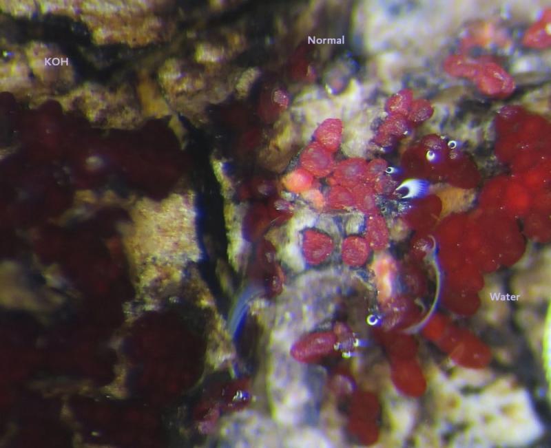
About the host, I currently have this pic. I try to get some spore images
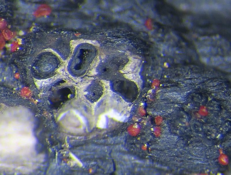
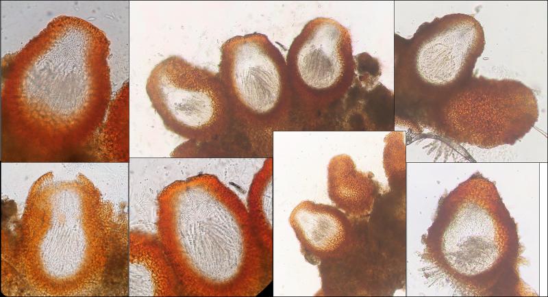
The perithecia seems to be fragiferiform-subpyriform, with a truncate apex. Wall warted (pustulate), bright red darkening at the top, paler below. When detached from the substrate, they measure about 0.23-0.29 mm in length and 0.19-0.23 in width (widest part). I couldn't produce a thin section!
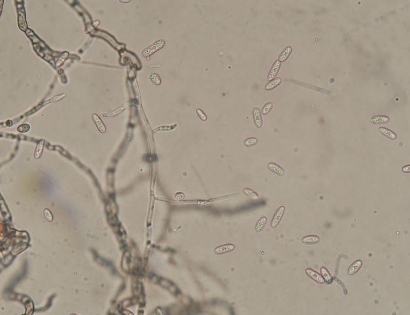
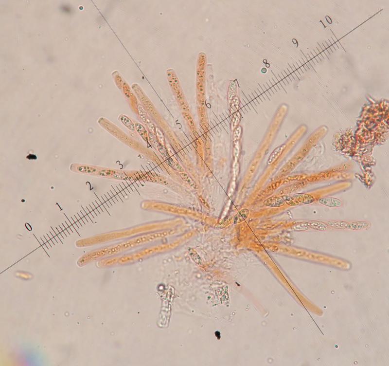
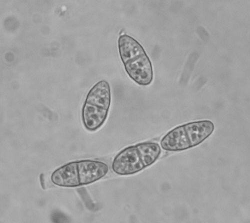
The ascii cylindrical, sides parallel (hence not swollen or fusiform) flexuose, with ascospores sitting at about 45 degrees stacked over each other in one series along most of the ascus length, apex truncate, J-ve, 75-95 um long by 6.5-8.5um wide
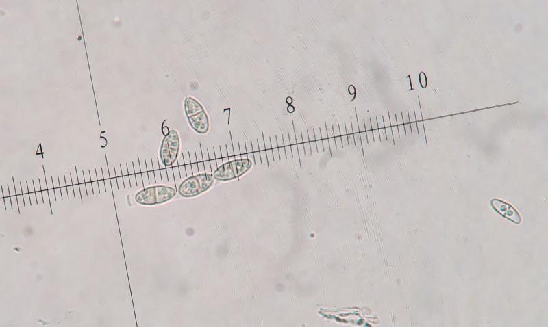
Ascospores
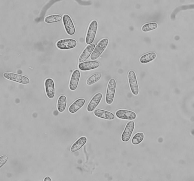
Navicular-fusiform, broadest at the middle, ends broadly rounded, one septate and slightly constricted at the septum, measuring (12.8) 13.8 - 17 (17.5) × (4.7) 5.6 - 6.9 (7.6) µm // Mean = 15.2 × 6.3 µm ; Qe = 2.5 ; Ve = 320 µm3


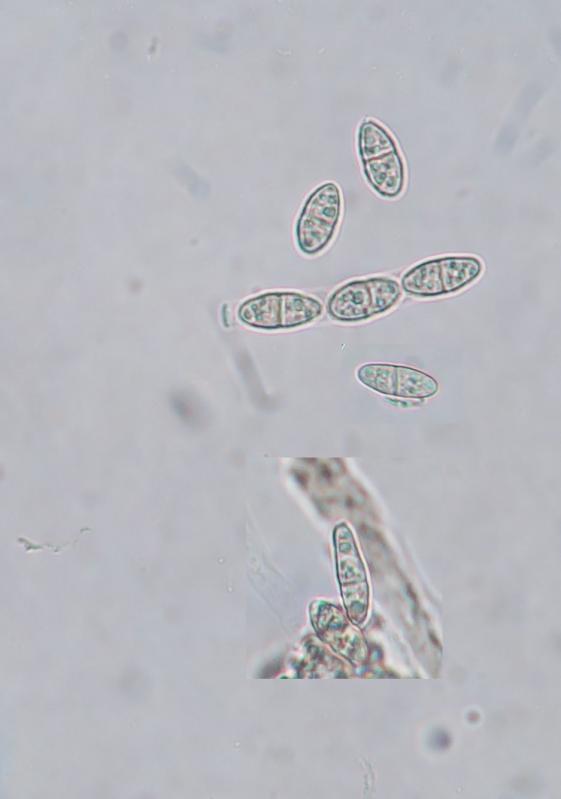
Here I attach images of cross sections of the ascomata


Thanks... your reply means that this is very strange! Some quick questions maybe you can put some light on this foggy picture:
1. Why have we abandoned Pseudcosmospora sp. ? Are the ascospores too large ?
2. Then, can it be a Cosmospora sp. or either that match with the genus concept?
3. Can you provide further help on the host (genus level)? Are those elongated spores typical of some family or genus?
I would love to learn what is unpeculiar with the data I presented.
I try to re-isolate, but the secimen is getting contaminated from airborne spores which makes life quie difficult
Cosmospora is characterised by an acremonium-like asexual morph in culture and green colony.
Pseudocosmospora also has an acremonium-like asexual morph, but rosy-buff, pale-luteous, or salmon-pink colony.
Cultures must be made in sterile environment, in laminar flow cabinet.
Good lucke
Christian
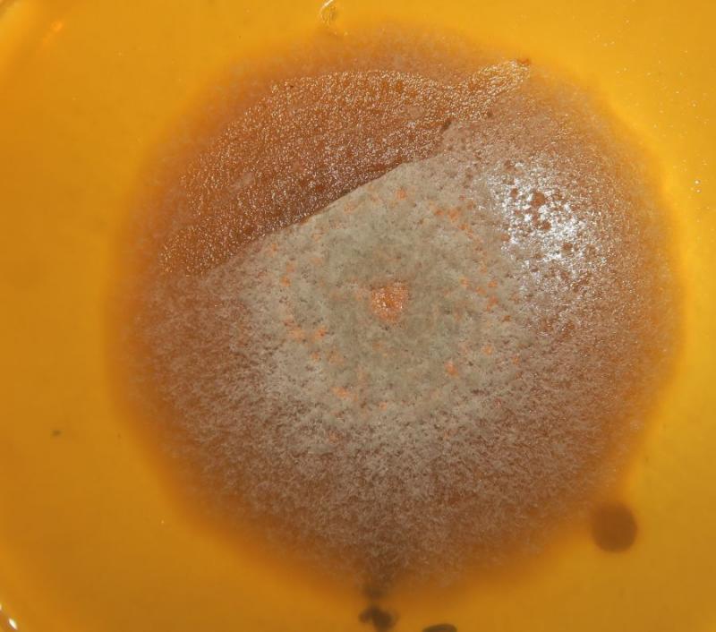
Acremonium-like anamorph and pale salmon colonies observed, but one colony had a a greenish hue when old. In one of the pics I think I got a pure colony (pinkish hue) and should make it a Pseudocosmospora but, yes not easy to draw a conclusion.
Ok, i think we have an interesting species and will progress with sequencing.
Many thanks...