
19-04-2026 21:23
Steve ClementsBonjour, I found this anamorphic fungus on old pl
19-04-2026 20:46
Steve Clements1 mm diameter approx spherical conidiophores on pl
12-04-2026 17:56
 Hardware Tony
Found on dead stems in February earlier this year
17-04-2026 19:16
 Enrique Rubio
Hi to everybodyI would appreciate any assistance r
14-04-2026 05:32
Ethan CrensonHi all, A few weeks back a friend pointed out som
17-04-2026 15:14
 Bruno Coué
Bonjour.Récoltes du 16/04/2026, sur feuilles mort
12-04-2026 15:52
Gernot FriebesHi,I'm looking for help with this anamorph collect
14-04-2026 21:52
Gernot FriebesHi,found on dead leaves of Carex elata. Conidia: 4
16-04-2026 22:09
Buckwheat PeteHello, I'd like to ask about this older specimen:
15-04-2026 19:33
Fátima Durán ManzanequeHi!! I need help, I found this Ascomycete but I d
 Hi,
Hi,Is this just a long spore variation of Hymenoscyphus scutula (H. aff. fucatus), or is it something else?
https://inaturalist.ca/observations/57610849
On rotten leaves, white when fresh and yellowish when dried.
The stalk base was white.
Spores:
(28.6) 29.5 - 36 (36.7) × (3.5) 3.9 - 4.4 (4.5) µm
Q = (6.6) 6.9 - 8.8 (9.9) ; N = 30
Me = 32.8 × 4.2 µm ; Qe = 7.9
Asci with a simple base, IKI+b.
Best regards,
Igor

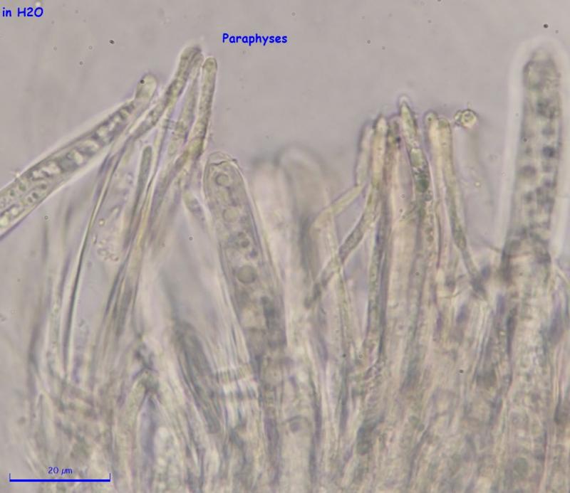
I did examine the specimen the next day, but this was more delicate than others and maybe it was too much for it. I took many pictures, but none of them shows the content of paraphyses well, but I attached the best what I have. It was my first year of hunting Hymenoscyphus like species and I'm lost in the process of recording and identification of them, so I'm not efficient, but I'm learning and maybe by the summer I will know what to do.
It is in an area where I hike a lot, so I will have a high chance of finding it again at the same spot.
Best regards,
Igor
Sorry for raising this topic again. I reexamined the specimen, looked at the textura and I think the medullary excipulum is porrecta and ectal excipulum is prismatica. I don't know if this helps. I uploaded the pictures (https://inaturalist.ca/observations/57610849).
I usually look at spores, paraphyses, and asci when they are still fresh and I take macro photos and I can do the rest later. What other steps should I do when I get fresh Hymenoscyphus-like specimen? Or what other tools should I be using?
Last year I was overwhelmed with a number of new species I was collecting. Usually, if I find something new it really slows me down and I was unable to look at all my specimens, so definitely I've missed a lot. This coming season I will revisit these places and I will get more fresh specimens. I want to be ready to handle them.
Thanks,
Igor
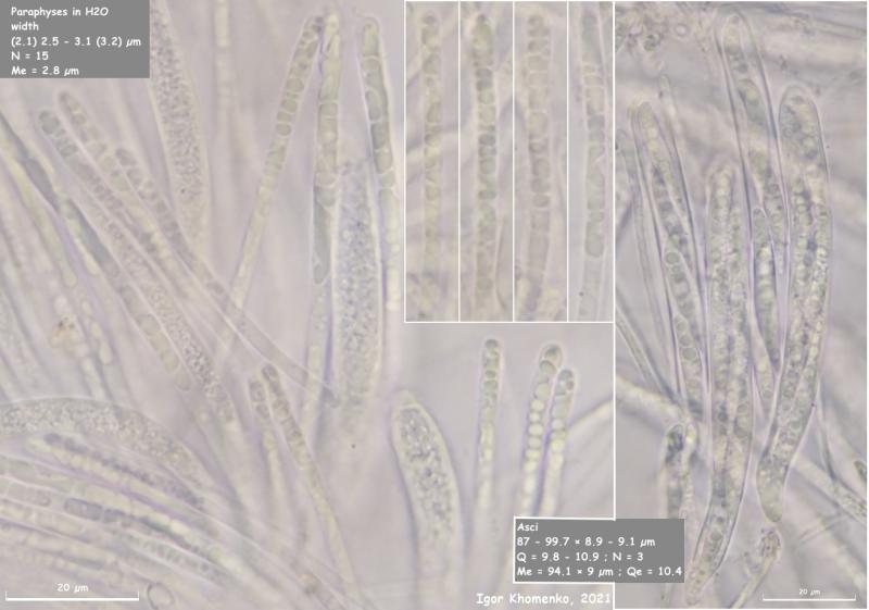
This Hymenoscyphus is back at the same spot as the last year and I collected enough to study.
This time I got a good picture of paraphyses. The spores slightly smaller ((26.7) 27 - 31.7 (35.6) × (4) 4.1 - 4.7 (4.9) µm), but the rest looks the same. I still don't know on what leaves it grows, but there is a chance that it could be Ulmus.
I looked for crystals in the stipe and couldn't find them, only saw cells with nucleus.
All new pictures are here:
https://inaturalist.org/observations/90185294
Igor
Regards,
Igor