08-06-2026 10:16
 Spooren Marco
I don`t have a clou about this fungus,it is not in
07-06-2026 15:10
 William Slosse
Hello everyone,On 05-06-26, I found following asco
08-06-2026 17:00
François BartholomeeusenGood day everyone, On June 5 2026, I collected de
05-06-2026 11:02
Thomas Læssøehttps://svampe.databasen.org/observations/10596691
07-06-2026 12:09
 François Freléchoux
Bonjour, Voici une brève description de ce qui m
07-06-2026 12:43
Steve ClementsBojour. This was a strange find on a stick on my
12-07-2015 00:05
 Nedim Jukic
This one from the same locality as the previous on
06-06-2026 17:44
Steve ClementsBonjour, This disco was on planed wood 3 x 1.5 cm
14-08-2016 23:15
 Alex Akulov
Dear friendsCan you help me to find the descriptio
Rutstroemia sp.
Perz Piotr,
23-02-2006 12:14
I have found this Rutstroemia on [unidetified] petiole: http://wwkk.mikologia.pl/spec/20051015_001_PERZ/20051015_001_PERZ.php
What is strange? Strange is H2O+IKI reaction on..... I do not know where :) It seems to be LB's in paraphyses reaction, or interhymenial gel (A.Gminder) ?? Something is here amyloid!
Rutstroemia sydowiana HAS NOT this reaction (H2O+IKI), may be Rutstroemia petiolorum, but i do not have this spec. in my herbarium and i can not test this reaction.
What do you think - is this a contaminant in hymenium or this taxon does have amyloid LB's in paraphyses OR interhymenial gel ?
Has anyone R.petiolorum and can make test for me? Or may be do you know this spec.?
Best,
Pimpek
Hans-Otto Baral,
23-03-2006 18:26

Re:Rutstroemia sp.
The Rutstroemia is very interesting, I think it is conspecific with R. "kalevi" on the DVD. This name is unpublished, it was intended to be published by J.T. Palmer and myself long ago. The substrate looks for me as an Acer petiole, and that´s also the substrate of the two earlier finds.
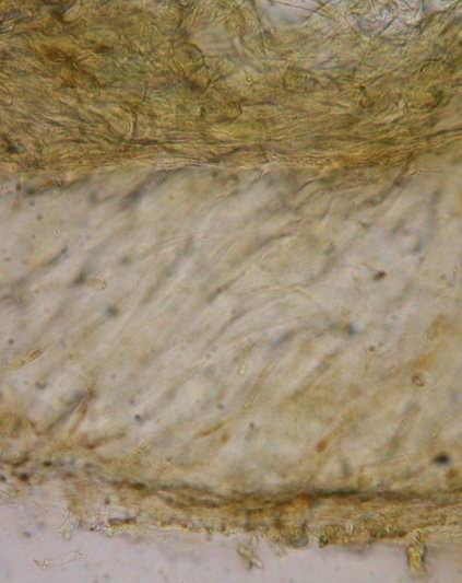
I found the ectal excipulum to be heavily gelatinized! No t. prismatica but strong oblita, see below. But it´s interesting: the find made by Lothar Krieglsteiner I saw fresh and I saw only scarcely any gel in the (living) excipulum, whereas in the find from Estonia the excipulum is heavily gelatinized. You write you have only one fruitbody, but I see two on your drawing. Do you still have one? Please have another look at the excipulum.
The species is surely close to R. coracina, but that is on Quercus leaves in the mediterranean area and has broader spores and brown vacuoles in the living paraphyses. Typical for both is the brown crenulate margin.
What I´m surprized from your drawing are the multiguttulate paraphyses. Such guttulation is very very typical of R. luteovirescens (which also grows on stromatized Acer leaves). But that species has yelow apothecia and really a textura prismatica without gel! R. kalevi has Mollisia-like vacuoles in the paraphyses. I presume that the guttules you saw in the dead material are secondary, maybe induced by the iodine. By the way I saw only the apical rings to react blue in IKI, and I am sure that you saw the blue rings of the old emptied and collapsed asci among the paraphyses and were misled to believe that the latter were amyloid.