
02-05-2026 12:42
Alain BRISSARDBonjour à tousJeuidi 30 avril dernier on m'a remi
02-05-2026 13:06
Pauline. PennaBonjour Please can someone help me with this id
01-05-2026 22:45
 Thierry Blondelle
Bonjour à tous, Une récolte sur bouse séchée d
28-04-2026 20:07
 Lothar Krieglsteiner
... on twig in the air at standing Ceratonia siliq
14-04-2026 05:32
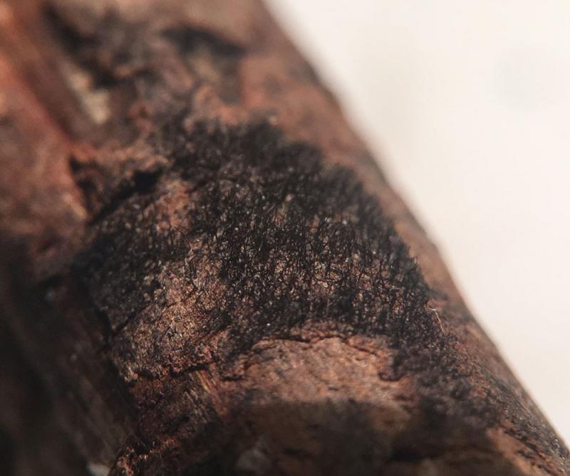
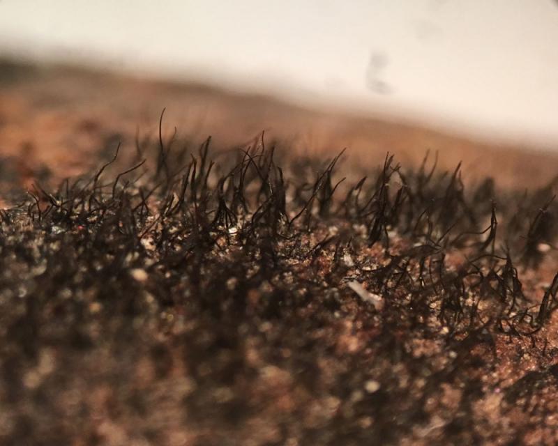
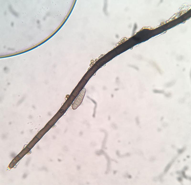
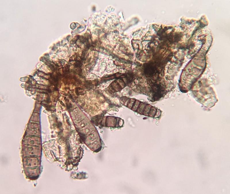
Ethan CrensonHi all, A few weeks back a friend pointed out som
28-04-2026 20:33
Vitus SchäfftleinHello, I found Trochila ilicina on Ilex aquifoliu

30-04-2026 10:28
Rot BojanHello, by appearance I would say that I am dealing
27-04-2026 18:48
Tony MoverleyCollected 23rd April 2026, Norfolk, EnglandSwarms
27-04-2026 20:52
Lothar Krieglsteiner
Found on hanging tiwg of Olea europaea in dried-ou
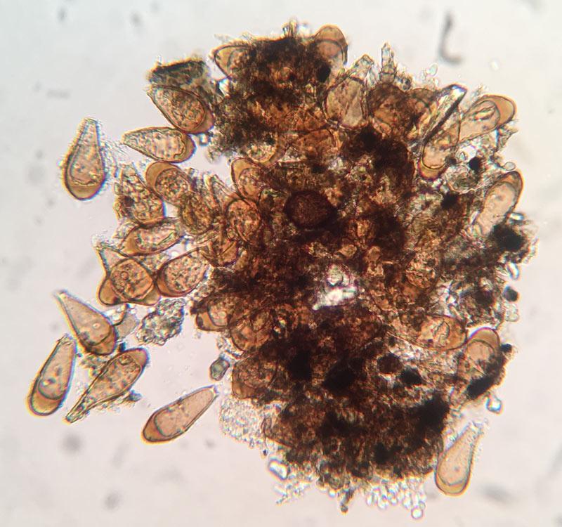
Associated with this mount I found two types of conidiospores, so I am uncertain which, if either, belongs to this organism...The first type: Shaped like a bowling pin (ellipsoid, but tapering to a narrow knob at one end), light brown, 4-8(+)septate, others simply ellipsoid, septate, about 32-72 by 10-15µm.
The second type of conidiospores I saw: Light brown, paddle shaped conidiospores with a thick wall but especially thick at the wide end, 35-55 by 17-22µm.
Any ideas? Thank you in advance!

Cheers!
Jason, again thank you!

Dear Ethan,
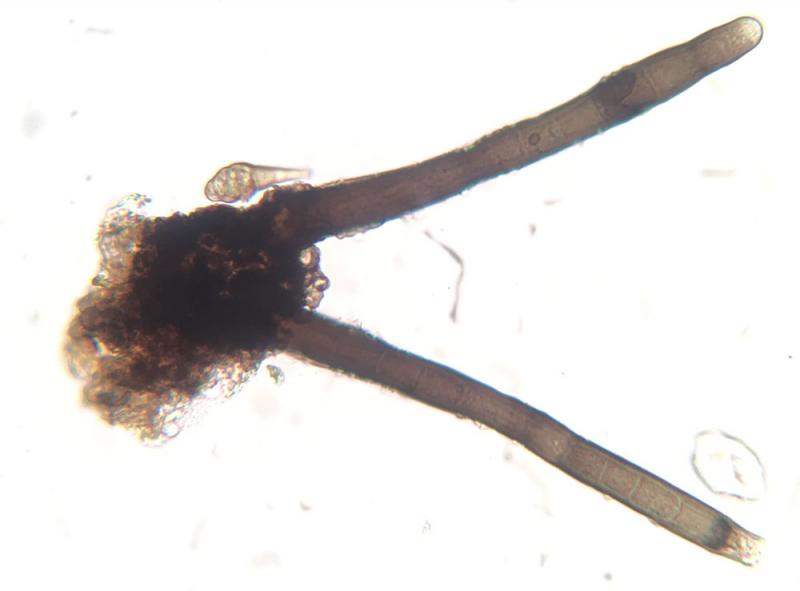
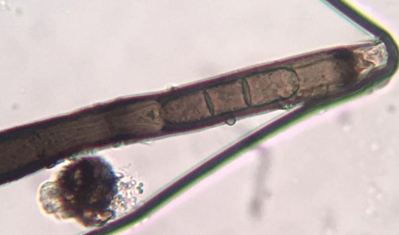
the hairy structures you illustrate match well conidiophores of Helminthosporium velutinum, which can form large effuse patches. In well preserved fresh specimens the acropleurogenous conidia sitting on the conidiophores are very distinct already under the hand lens, but your material may be rather old, so the conidia have already fallen off, or they didn't yet produce conidia (diffficult to decide without detailed microscopic investigations). In one of your pictures, these typical Helminthosporium conidia are well seen. As remains of spore production, in the conidiophores you should observe marginal pores in the upper cells, and also one at the top. However, sometimes many conidiophores remain sterile if the conditions for conidiation are unfavourable. Conidiophores can also stop growth and grow out again, this is also seen in some of your pictures.
See our publication on Helminthosporium in Studies in Mycology (freely downloadable at https://www.sciencedirect.com/science/article/pii/S0166061617300210)
There are also other Helminthosporium species around, for which you need more detailed investigations and measurements of conidiophores and conidia, but the most common species in temperate areas seems to be Helminthosporium velutinum. This species is also polyphagous and known from many different mainly woody but sometimes also herbaceous hosts.
The other type of conidia may represent undeveloped Helminthosporium conidia, this sometimes happens when there are unfavourable conditions during conidiogenesis and the conida die off before they reach full maturity. The conidia illustrated are obviously already dead. As the septa of the conidia of Helminthosporium are distosepta, they fully degrade some time after death, and only the outer wall is left over, which leaves the impression that the conidia are aseptate.
Cheers,
Hermann
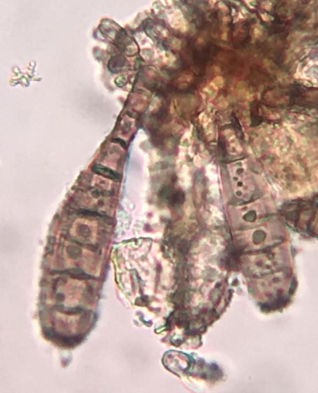
Hi Jason,
what you identify as conidia are in fact conidiophores which terminated growth and resumed growth again at the apex. The same can happen when the tip is damaged, eaten by an insect etc, then the conidiophore resumes growth, leading to this phenomenon. Can be sometimes observed in Helminthosporium. The tip of the lower condidiophore is actively growing, but due to high contrast the outline of the hyaline wall there not fully visible.