
29-05-2026 15:35
daniel FERREBonjour à tous,Je voudrais votre aide pour cette
28-05-2026 16:15
James MitchellHello,Does anyone have the original publication of
28-05-2026 11:06
Thomas Læssøehttps://svampe.databasen.org/observations/10596750
23-05-2026 11:44
 Charles Grapinet
Hello, I am having trouble identifying this copro
25-05-2026 16:44
François BartholomeeusenHi forum members,During an excursion organised by
26-05-2026 21:25
Dirk GerstnerHello everyone, I'm completely stumped by this li
26-05-2026 22:44
Ethan CrensonHi all, I think I have Incrucipulum capitatum her
22-05-2026 14:44
 Lothar Krieglsteiner
in unripe condition citrine yellow, then soon fadi
25-05-2026 16:35
 Bernard CLESSE
Bonjour à toutes et tous,J'ai trouvé récemment,
22-05-2026 13:29
Gernot FriebesHi,I am curious to hear your opinion on this mater
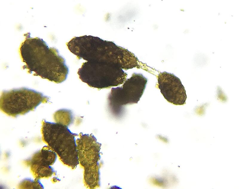
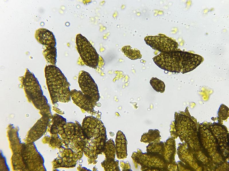
Berkleasmium conglobatum (?)
Ethan Crenson,
11-08-2017 17:19

Hans-Otto Baral,
11-08-2017 19:14

Re : Berkleasmium conglobatum (?)
I have seen a similar fungus on dead wood of Acacia in arid Australia, but the conidia were max. 30 µm long. I noticed in this species a strong ionomidotic reaction of the conidia in KOH (orange stain extruding in the medium). Did you test that?
Zotto
Zotto
Jason Karakehian,
11-08-2017 19:50

Re : Berkleasmium conglobatum (?)
Hi Ethan, I posted this species to our Facebook group in June and I just sent you a message with the link to that post. Here is a link to my post in Mycoportal:
http://mycoportal.org/portal/collections/individual/index.php?occid=4622329
I think your determination is correct. The conidia seem to darken in age to nearly opaque black. The farinaceous or flaky condition of the surface of the conidia is consistent with my observations. Also, you will see nearly black sporodochia in a collection and also these yellow-green sporodochia. These are younger sporodochia that have had the tops rubbed away and you see this yellow tissue (hyphae and conidiogenous cells) beneath. Best - Jason