
19-04-2026 21:23
Steve ClementsBonjour, I found this anamorphic fungus on old pl
19-04-2026 20:46
Steve Clements1 mm diameter approx spherical conidiophores on pl
12-04-2026 17:56
 Hardware Tony
Found on dead stems in February earlier this year
17-04-2026 19:16
 Enrique Rubio
Hi to everybodyI would appreciate any assistance r
14-04-2026 05:32
Ethan CrensonHi all, A few weeks back a friend pointed out som
17-04-2026 15:14
 Bruno Coué
Bonjour.Récoltes du 16/04/2026, sur feuilles mort
12-04-2026 15:52
Gernot FriebesHi,I'm looking for help with this anamorph collect
14-04-2026 21:52
Gernot FriebesHi,found on dead leaves of Carex elata. Conidia: 4
16-04-2026 22:09
Buckwheat PeteHello, I'd like to ask about this older specimen:
15-04-2026 19:33
Fátima Durán ManzanequeHi!! I need help, I found this Ascomycete but I d
 Hi,
Hi,I (totally inexperienced) would kindly ask for some help. Forgive me eventually stupid ideas.
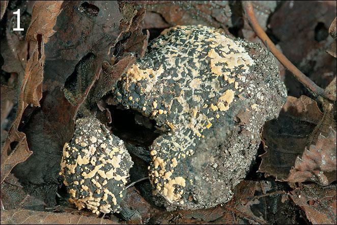
I found this tuber (hope so) with what I believe is a parasitic fungus on it. The fruitbody seemed to be slightly over-matured. Originally it was a single body, but it broke into two pieces during handling (Fig.1).
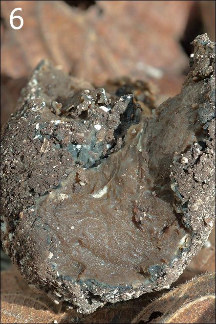
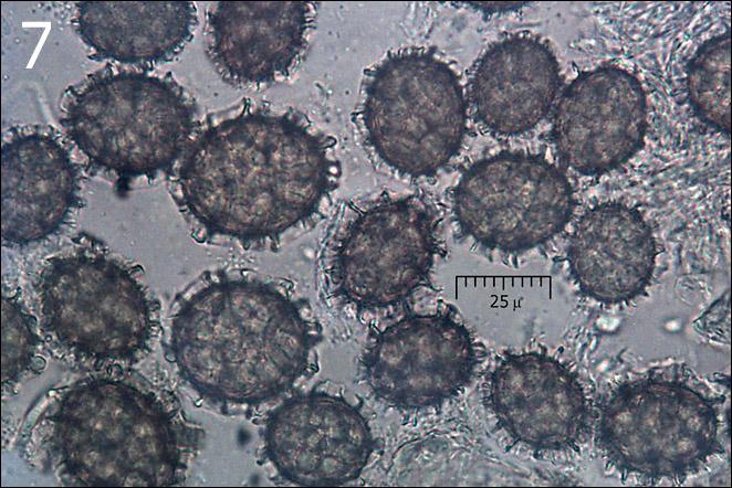
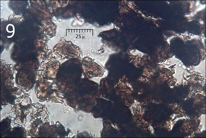
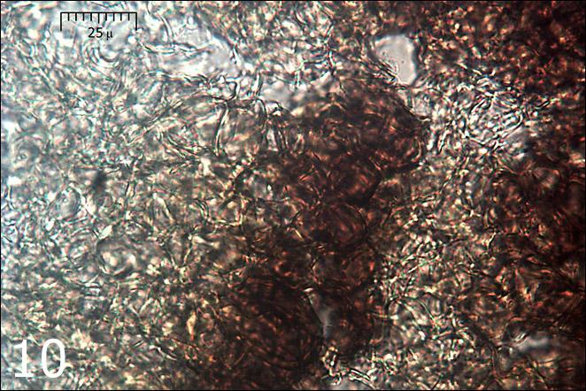
Based on spore dimensions (Me = 26 x 20.8 micr., n= 51) (Fig.7,8), their coarsely reticulated surface and tuber's cortex it could eventually be Tuber aestivum/uncinatum (Figs. 1-6). But the date of the find is very late, on November 3, and cross-section doesn't fit to what I can find in literature and internet (over-mature tuber?). It also had very faint smell. Also, it was not really buried in the ground. Gleba was almost 100% spore mass. I found only a few places where I believed I saw 'remnants' of asci (Fig.8, arrow). Variation in spore dimensions was enormous (Fig.8, from 21 to 45 micr. diameter). Tissue below cortex (Fig. 9, 10) was interesting, like a spatial 'hexangular' net (Fig.10) with no spores there. Can this really be Tuber aestivum?
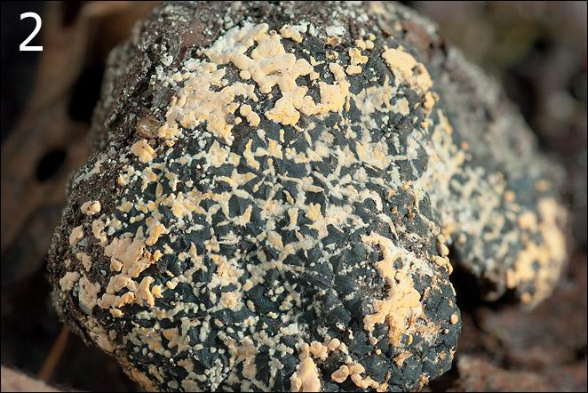
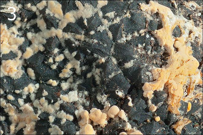
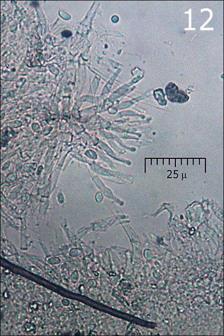
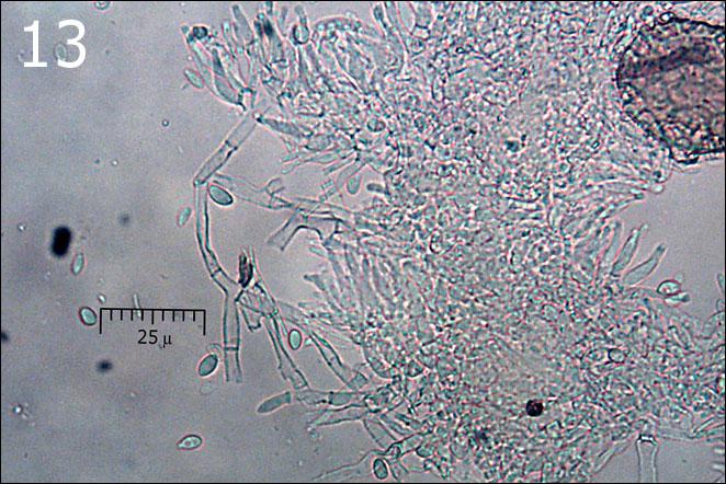
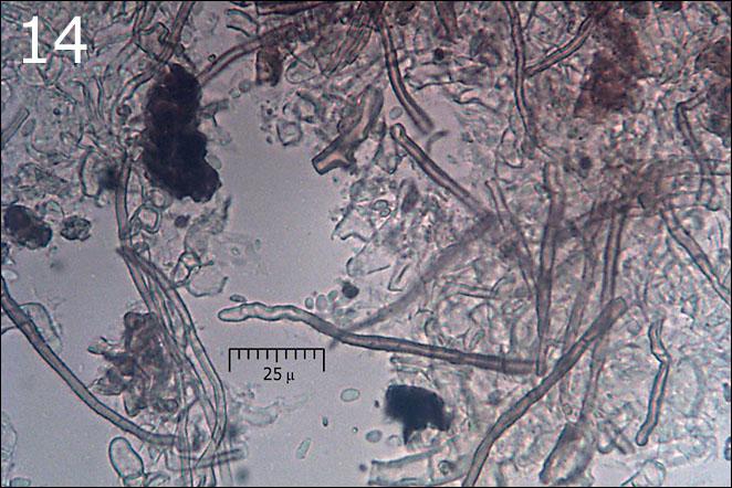
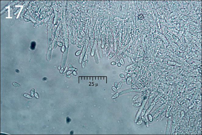
Orange blobs on tuber are probably a parasitic fungus, eventually an anamorph (Figs. 11-14) (Sympodiophora / Hypomyces ??). I've found neither asci nor basidia. Hypha is of two kinds, thin walled (Fig. 13) and yellow-brown thick walled (Fig.14). No clamps seen. Squashes of the blobs were full of small, smooth spores/conidia (Me = 5.7 x 3.4 micr., n = 40) shown on Fig.15. The surface of the blobs show a kind of flask shaped 'cystidia' with globose apex (Fig.12) - possibly conidiophores?
On the lower surface of the tuber I also noticed a few small (fi 1-2 mm) circular, cottony tufts of snow white hypha which seems to be a kind of mold (old tuber?). Under microscope I found only hypha without any other structure (Fig. 17) in them.
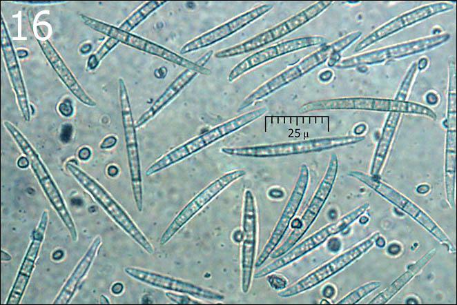
A third type of spores/conidia was also observed (Fig. 16) - very numerous, long and narrow, spindle shaped and septated (Me = 49.3 x 5.3 micr, n = 31; 3 - 7 septa, avg = 4.9, n = 35). They were everywhere, but seem to be associated mostly with the tuber's gleba (not sure). Where they actually belong I don't know.
All three types of spores/conidia were very numerous, so I exclude the possibility of an external contamination of the find.
I will be grateful to anybody for shedding some light on this interesting puzzle (for me).
Regards
Amadej
______________________
Habitat: mountain slope; Fagus sylvatica forest with some Picea abies; 590 m asl; in shade; colluvial, skeletal, calcareous ground; average temperature 7-9 deg C, average precipitations about 3.000 mm/year; alpine phytogeographical region.
Substratum: apparently on soil (not in!), but almost completely buried with ground humus, mostly rotten leaves of Fagus sylvatica.
Place: Trenta valley, East Julian Alps, Slovenia.
______________________


Best regards Walter
Warmest regards
Amadej
Please send it air-dry and not air-tight closed, because I shall be absent for 3 weeks.