
12-06-2026 14:50
 François Freléchoux
Bonjour, Voici la brève description d'une Mollis
10-06-2026 21:16
François Freléchoux
Bonsoir,Le dernier du jour, en attendant votre avi
11-06-2026 19:01
 William Slosse
Hello all,In an attempt to make a culture of a sus
11-06-2026 19:03
 Nicolas VAN VOOREN
Chers membres d'Ascofrance,Le site sera placé en

09-06-2026 18:32
Camille MertensSur morceau de roseau immergé 0,5 - 0,7 mm de dia
10-06-2026 12:54
Steve ClementsBonjour encore, Pouvez-vous m'aider, s'il vous pl
10-06-2026 21:07
François Freléchoux
Toutes les tiges de gentianes jaunes de l'an pass�
10-06-2026 13:41
François Freléchoux
Bonjour à nouveau, Voici une trouvaille d'hier.
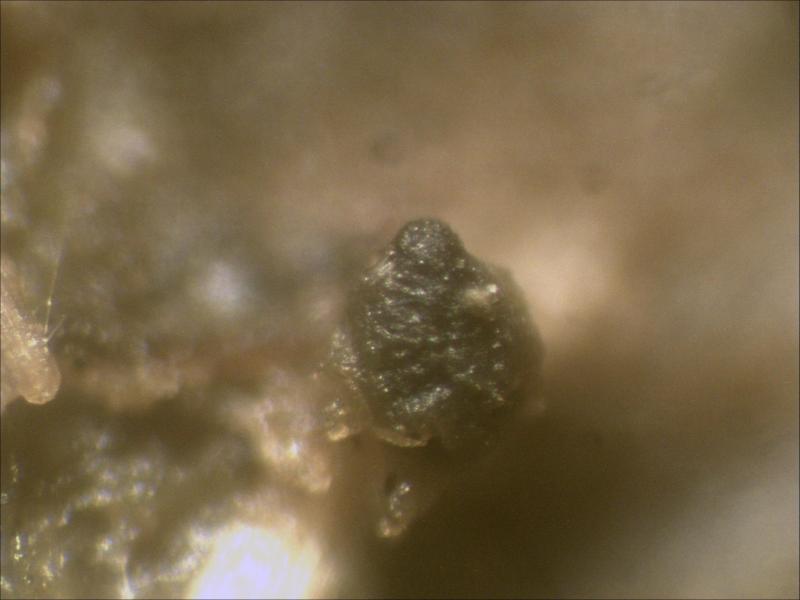
Sporormiella heptamera
Joop van der Lee,
29-11-2015 12:57
 I did find Sporormiella heptamera on cow dung and at the same time observed some items I cannot find in documentations.
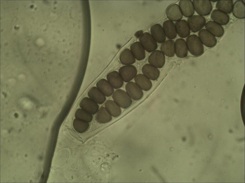
Asci when within the fruitbody 234.7x59.7 um, seperately attached stipe 50.3x10.5 um with long end 29.2 um. Ascus containing eight 7-celled spores. Spores are tri-serial in the upper and middle part en bi-serial in the lower part.
As soon as the asci leave the fruitbody and come in contact with water they will expand measuring approx. 369x52.6 um with attached stipe 67x22.3 um and long end of 79 um
The outer wall of the ascus is rolled up near the attachment of the stipe and only the innner part of the ascus remains 21-21.5 um containig the spores.
Young spore elements are rounded and fully attached to each other the total length is 79.5-85.5x17-18.8 um (for measurement of the width I only used the 3rd cell from the upper).
Half mature spores: still fully attached to each other, bended 72.3-75.5x17.6-20.0 um.
Mature spores: not fully attached 90.5-92.0x19-21.46 um, bended, spores are covered with a gelatineus sheath.
Germs slit on the middle cell elements run on both sides the jump from one side to the other is made by a sinousodial transverse slit. The total slit ends for both sides when it reaches the transvers slit.
For the end cells the slit runs downwards around the conical top.
The end cells are nearly equal in size for the width but the lower end cell is longer than the upper.
The 3rd cell from the upper is the greatest while the others are gradually decrease in size towards the lower end cell.


Peter Püwert,
29-11-2015 13:26

Re : Sporormiella heptamera
Hi Joop,
the done observations are already interesting, but also explicable. Asci extend in the water because the pressure rises inside bitunicate asci. The middle separate segments of the spores have a similar form like Coniochaeta spores, lenticular. I hope to have understood the questions correctly.
Greetings
Peter.
Joop van der Lee,
29-11-2015 13:39
Re : Sporormiella heptamera
Thanks for your comment Peter but Cain mentioned in his discription of S. heptamera that the ascus is contracted below into a slender stipe but here that is not the case nor is there any notice of the fact that the outer layer of the ascus is rolled up near the conntection of the broader part of the stipe with the ascus.
And secondly I could not find that the germ slit runs all the way round on both sides of the spore cells ending just before the transvers slit.
And secondly I could not find that the germ slit runs all the way round on both sides of the spore cells ending just before the transvers slit.
Peter Püwert,
29-11-2015 15:03
Re : Sporormiella heptamera
Hi Joop,
pulling together the exterior cover in this point is definitely no support for the determination but pure coincidence. With germ slits I do not understand or the translation suffers by the special terms.
Greetings Peter.
pulling together the exterior cover in this point is definitely no support for the determination but pure coincidence. With germ slits I do not understand or the translation suffers by the special terms.
Greetings Peter.