
30-05-2026 21:12
Philippe PELLICIERSur branche de mélèze (Larix) près de la neige,
25-05-2026 16:35
 Bernard CLESSE
Bonjour à toutes et tous,J'ai trouvé récemment,
29-05-2026 15:35
daniel FERREBonjour à tous,Je voudrais votre aide pour cette
28-05-2026 16:15
James MitchellHello,Does anyone have the original publication of
28-05-2026 11:06
Thomas Læssøehttps://svampe.databasen.org/observations/10596750
23-05-2026 11:44
 Charles Grapinet
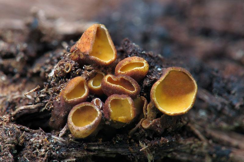
Hello, I am having trouble identifying this copro
25-05-2026 16:44
François BartholomeeusenHi forum members,During an excursion organised by
26-05-2026 21:25
Dirk GerstnerHello everyone, I'm completely stumped by this li
26-05-2026 22:44
Ethan CrensonHi all, I think I have Incrucipulum capitatum her
22-05-2026 14:44
 Lothar Krieglsteiner
in unripe condition citrine yellow, then soon fadi
Encoelia ??
Leandro Sánchez,
04-05-2015 11:21
 Sur feuillus, diamètre max 4 mm.
Asques 43-52 / 5-6 , croziers +, IKI -
Je pense Encoelia fimbriata mais je n'ai information, je cherche cet article
Encoelia fimbriata Spooner & Trigaux, Trans. Br. mycol. Soc. 85(3): 547 (1985)
Merci d'avance.
Cordialement
Gilbert MOYNE,
04-05-2015 12:11
Re : Encoelia ??
Je serais aussi intéressé par cet article. Merci.
Ici, je serais curieux de connaître la réaction de la chair à KOH car cela me fait un peu penser à Ionomidotis fulvotingens;
Gilbert
Ici, je serais curieux de connaître la réaction de la chair à KOH car cela me fait un peu penser à Ionomidotis fulvotingens;
Gilbert
Gernot Friebes,
04-05-2015 12:42
Re : Encoelia ??
Here's a link to the article: http://www.cybertruffle.org.uk/cyberliber/59351/0085/003/0547.htm
Best wishes,
Gernot
Best wishes,
Gernot
Leandro Sánchez,
04-05-2015 13:11
Re : Encoelia ??
Thank you very much !!!!!
Leandro Sánchez,
04-05-2015 15:24
Re : Encoelia ??
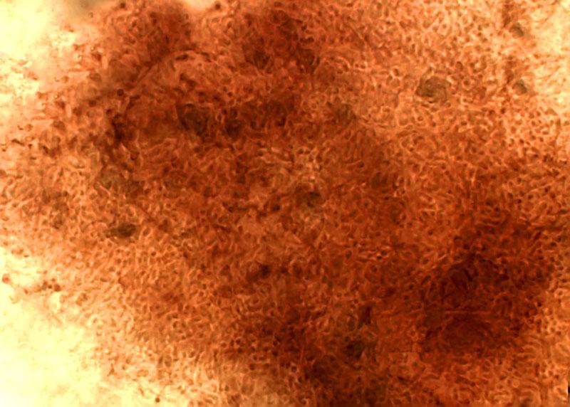
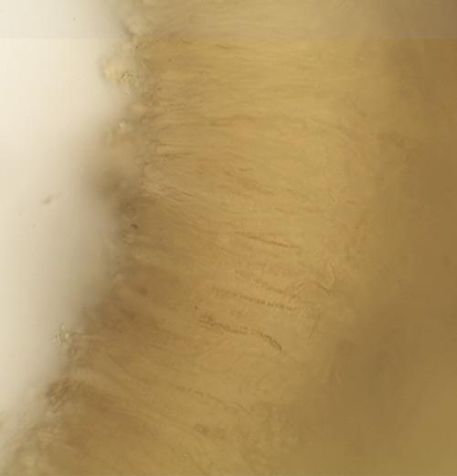
Je n'ai observé poils, photo chair KOH 5 %
Cordialement
Cordialement
Gilbert MOYNE,
04-05-2015 15:38
Re : Encoelia ??
Donc pas Ionomidotis...
Un Encoelia donc mais lequel si pas de poils ?
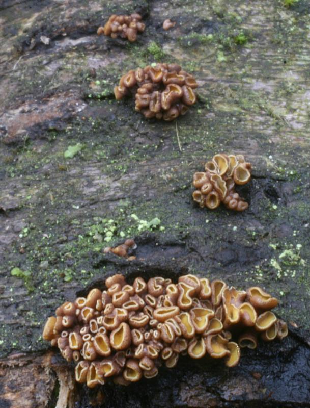
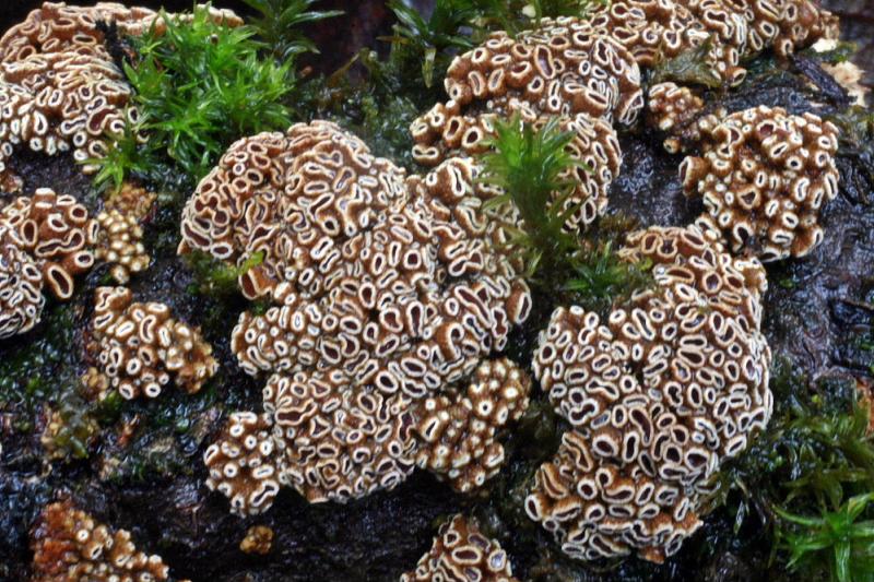
Voici une photo de Encoelia fimbriata. C'est assez ressemblant mais les apothécies sont en général beaucoup plus serrées.
Gilbert
Un Encoelia donc mais lequel si pas de poils ?
Voici une photo de Encoelia fimbriata. C'est assez ressemblant mais les apothécies sont en général beaucoup plus serrées.
Gilbert
Hans-Otto Baral,
05-05-2015 10:00

Re : Encoelia ??
Hello together
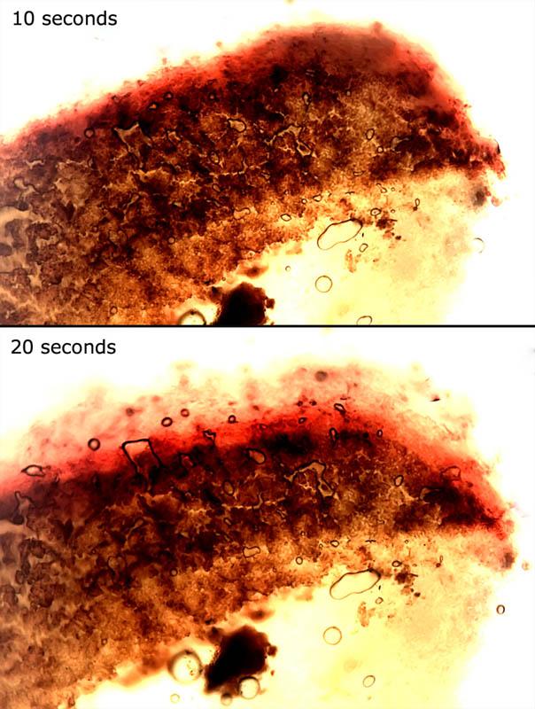
Your two collections look very interesting and at first sight I would estimate they are the same. I assume that the ionomidotic reaction was positive in both. Leandro, did you look for the moment when the KOH came in contact with the fungus? Only then you see for some 10-20 seconds the reaction, i.e. the dissolution of yellow or red-brown pigment.
E. fimbriata is a very special fungus, it has a complex internal texture (in the stipe region), with large amounts of crystals. Gilbert, your fungus does not look much hairy. Do you have a microanalysis?
The photos do not really remind me of Ionomidotis fulvotingens s.l.
Zotto
Leandro Sánchez,
05-05-2015 16:27
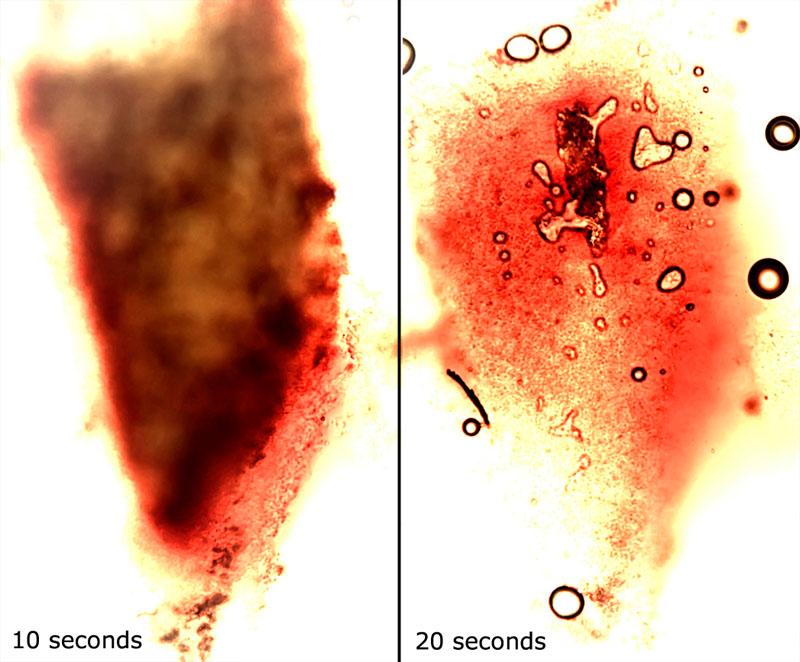
Re : Encoelia ??
The photos of the same area, with KOH 5 %, seconds after.
Hans-Otto Baral,
05-05-2015 16:47
Re : Encoelia ??
Yes, it is not very clear but your first pic on the right looks like if a red-brown pigment has extruded. Perhaps you should use a shorter shutter speed, I assume the area around was not white.
This is perhaps the ochraceous form of Ionomidotis fulvotingens, which is obviously a separate species, genetically quite distant from the olivaceous-blackish one. And maybe Gilbert has the same species, not fimbriata.
You do not know the substrate, what could it be, what kind of forest did you collect?
Gilbert MOYNE,
05-05-2015 17:42
Re : Encoelia ??
Bonjour Zotto et Lendro
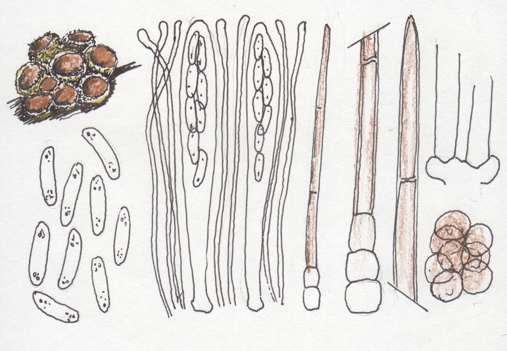
Une autre photo de Encoelia fimbriata et ma fiche micro de la récolte. Sur la première photo, je ne me souviens pas mais sur la deuxième, les poils étaient bien présents.
Amitiés
Gilbert
Une autre photo de Encoelia fimbriata et ma fiche micro de la récolte. Sur la première photo, je ne me souviens pas mais sur la deuxième, les poils étaient bien présents.
Amitiés
Gilbert
 Encoelia-fimbriata-11-0001.jpg
Leandro Sánchez,
05-05-2015 17:51
Re : Encoelia ??
River forest, but i dont know, perhaps Fraxinus, Alnus, etc
Hans-Otto Baral,
05-05-2015 17:55
Re : Encoelia ??
Yes, Gilbert, this is E. fimbriata. But the other not. This other one has indeed at the margin some subhyaline hairs as well. Was it also on Salix, and did you keep the specimen?
Hans-Otto Baral,
05-05-2015 18:02
Re : Encoelia ??
Fraxinus would be easy to recognize: it is a ring-pored wood.
If you want you can send this specimen, then I would try to confirm identity. I am not sure if Kadri Pärtel wants to sequence it. I will ask her.
If you want you can send this specimen, then I would try to confirm identity. I am not sure if Kadri Pärtel wants to sequence it. I will ask her.
Gilbert MOYNE,
05-05-2015 18:10
Re : Encoelia ??
Oui Zotto, sur Salix.automne hiver.
Pas gardé, j'avais dû envoyer à Michel Hairaud.
mais je dois pouvoir la retrouver l'hiver prochain sur le Marais de Saône, une zone humide proche de chez moi d'où elle provenait.
Gilbert
Pas gardé, j'avais dû envoyer à Michel Hairaud.
mais je dois pouvoir la retrouver l'hiver prochain sur le Marais de Saône, une zone humide proche de chez moi d'où elle provenait.
Gilbert
Leandro Sánchez,
05-05-2015 19:40
Re : Encoelia ??
Ok Zotto, I will send this specimen.
Thank you Zotto and Gilbert
Thank you Zotto and Gilbert
Hans-Otto Baral,
06-05-2015 11:17
Re : Encoelia ??
Thanks! So please address it as follows:
Hans-Otto Baral
Blaihofstr. 42
D-72074 Tübingen
Germany
Hans-Otto Baral
Blaihofstr. 42
D-72074 Tübingen
Germany
Hans-Otto Baral,
19-05-2015 11:25
Re : Encoelia ??
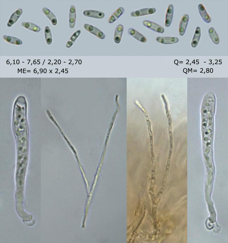
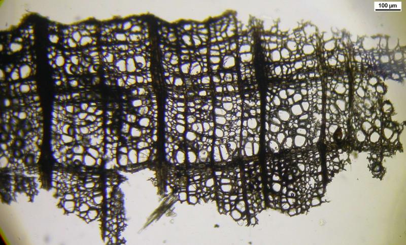
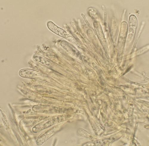
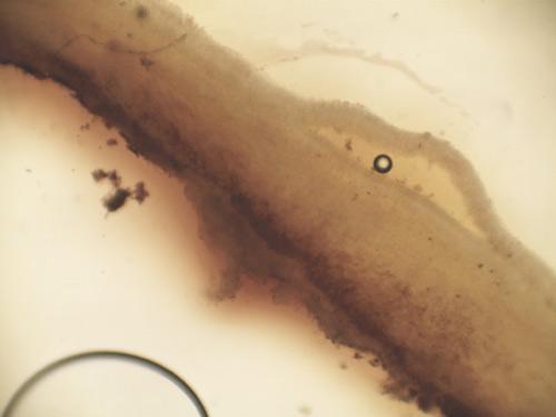
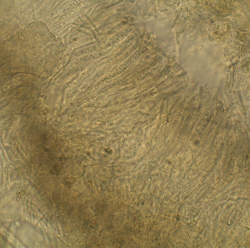
Yesterday I received the fungus. Here a few pics. I found the spores a bit smaller: *(5-)5.5-6.5(-7) x (1.7-)1.9-2.1 µm.
The asci have a prominent apical thickening in the dead state, especially when immature.
The yellow pigment in the flesh is striking, and also its solubility in KOH.
Now to the substrate : it is clearly Tilia. A small portion of bark looked characteristic, as does the wood anatomy.
We have no clear name yet for this fungus, perhaps "Cenangium" alnicola.
Please let me know the locality and further data (date, collector). HJow thick was the branch?
Zotto

Leandro Sánchez,
19-05-2015 16:02
Re : Encoelia ??
Thank you very much Zotto.
31T 430059.08 mE 4668868.86 mN
Les Lloses (Girona) 746 msnm
Javier Bometón, Leandro Sánchez & José Luis Martín
branch 40 mm Ø
Best regards
31T 430059.08 mE 4668868.86 mN
Les Lloses (Girona) 746 msnm
Javier Bometón, Leandro Sánchez & José Luis Martín
branch 40 mm Ø
Best regards
Hans-Otto Baral,
19-05-2015 17:08
Re : Encoelia ??
Thanks! With Google earth I arrive at 4.5 km SW of Ripoll, 742 m alt.
What was the date? Did you keep part in a herbarium, which number?
What was the date? Did you keep part in a herbarium, which number?
Leandro Sánchez,
19-05-2015 17:20
Re : Encoelia ??
Sorry, I forgot
01-05-2015
herbarium LSS 20150501-1
Best regards
01-05-2015
herbarium LSS 20150501-1
Best regards
Hans-Otto Baral,
19-05-2015 17:55
Re : Encoelia ??
Thanks!
I still wonder about the red versus yellow colour in KOH, does it originate from the camera, or did you see a red colour through the microscope?
I still wonder about the red versus yellow colour in KOH, does it originate from the camera, or did you see a red colour through the microscope?
Leandro Sánchez,
19-05-2015 19:04
Re : Encoelia ??
I saw clearly a red color in the microscope when entering the koh, i also think Javier
Hans-Otto Baral,
19-05-2015 20:09
Re : Encoelia ??
This is quite strange because that would be typical of Ionomidotis fulvotingens. But maybe it is more a question of the thickness of the preparation.
Bometon Javier,
19-05-2015 21:33

Re : Encoelia ??
Hola Zotto, Como dice Leandro, la pigmentación amarilla de la carne es claramente roja con KOH a través del ocular del microscopio. También rapidamente es soluble.
Saludos
Javier
Saludos
Javier
Hans-Otto Baral,
19-05-2015 21:43
Re : Encoelia ??
Yes, but on your photos no yellow at all is visible, although the fungus has a yellow flesh. I thought that your camera suppresses this? Is it a microscope camera without the possibility to manipulate the colour balance?
Bometon Javier,
19-05-2015 22:59
Re : Encoelia ??
Hola Zotto, el la foto queria mostrarle las ascas vivas y el rojo, seguro que con la cámara fotográfica salen mejor los colores que con la del microscopio, en las fotos que envío se ve algo mejor el amarillo.
Saludos
Javier
Saludos
Javier
Hans-Otto Baral,
20-05-2015 08:43
Re : Encoelia ??
Hi Javier
The photo with living asci is very good. Do you have it in higher resolution and with a scale?
The amarillo I do not see on my screen :-(
This part I do not understand, the translator produces nonsens: seguro que con la cámara fotográfica salen mejor los colores que con la del microscopio. - Ah, I think it means: a normal camera produces better colours than a microscope camera. - With the normal the problem is also the automatic white balance, but you can chose other options there, what seems to be impossible with microscope cameras.
Zotto
Bometon Javier,
21-05-2015 21:39
Re : Encoelia ??
Hola Zotto, le envío foto con mayor resolucción.
Si, es lo que quería decir, que se consigue mayor calidad con la cámara fotográfica
Gracias
Javier
Si, es lo que quería decir, que se consigue mayor calidad con la cámara fotográfica
Gracias
Javier