14-08-2016 23:15
 Alex Akulov
Dear friendsCan you help me to find the descriptio

05-06-2026 11:02
Thomas Læssøehttps://svampe.databasen.org/observations/10596691
04-06-2026 11:36
Gernot FriebesHi,found on Vaccinium myrtillus.Asci: IKI –, 8-s
05-06-2026 12:10
 François Freléchoux
Capitotricha sp. sur Lonicea caerulea Caractères
19-05-2026 10:27
Patrice TANCHAUDBonjour, récolte récente sur terre retournée i
04-06-2026 18:39
Gernot FriebesHi,I collected this species in two different locat
22-05-2026 13:29
Gernot FriebesHi,I am curious to hear your opinion on this mater
Bonjour !
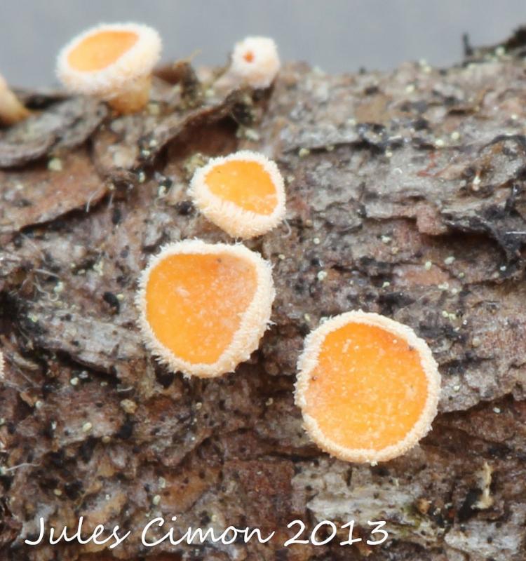
Voici un Lachnellula willbommii probable.
La pousse sur mélèze et les grandes spores sont conformes.
Merci de nous aider !
Roland
Données :
Date de récolte : 22 / 06 / 2013
Substrat : branche morte de mélèse
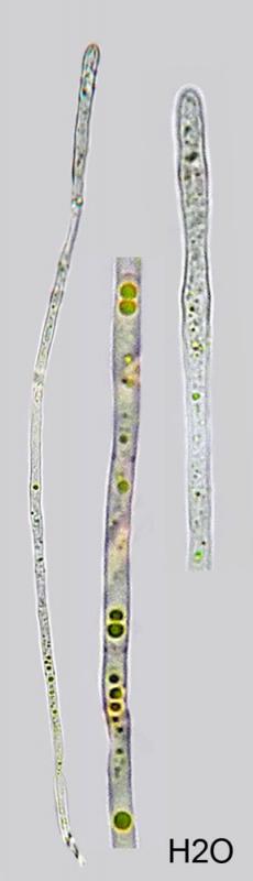
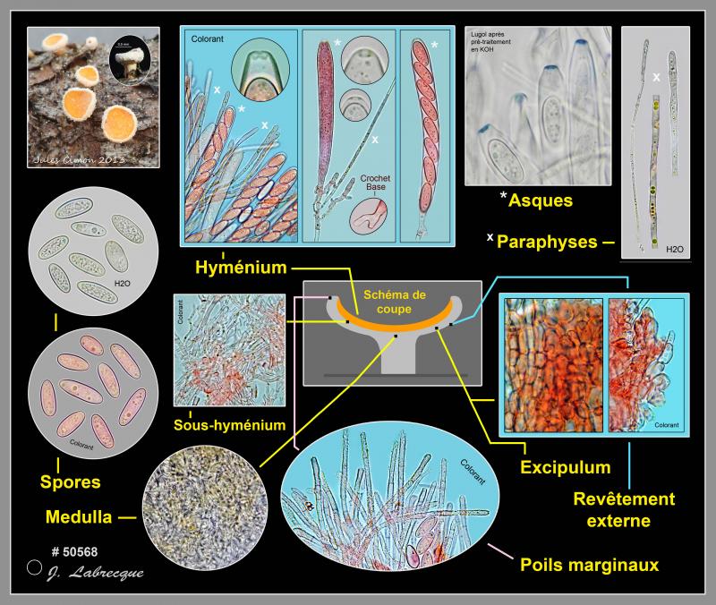
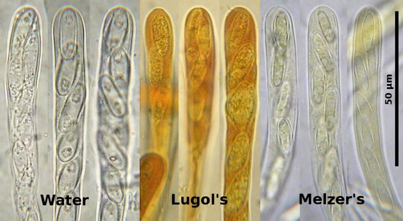
Asques non operculés, à 8 spores unisériées, avec crochets à la base et pore apical fortement amyloïde avec prétraitement dans le KOH puis dans le Melzer, à contenu légèrement dextrinoïde vers l'apex, 110-127 x 9-12 µm
Paraphyses étroitement cylindriques, parfois légèrement plus larges vers l'apex et avec un septum vers la base, non ramifiées, à contenu lipidique et fines guttules jaunes réfringen-tes jusqu'à 80% et plus, 113-170 x 2,5-4 µm, dépassant les asques d'environ 5-15 µm
Spores moyennement à étroitement ellipsoïdes, rarement subfusoïdes, lisses, à paroi légèrement épaissie, avec 1-2 guttules, 12-20,5 x 5,5-7,5 µm, 15,7 x 6,4 µm en moyenne, Q = 2,45
Sous-hyménium formé de petites hyphes emmêlées, sinueuses, septées, 1,5-2 µm de diam.
Excipulum en textura prismatica ascendant, gélatinisé, formé d'hyphes septées, à paroi épaissie, 2-3 µm de diam.
Revêtement externe formé de cellules globuleuses à ellipsoïdes, à paroi épaissie jusqu'à 1 µm de largeur, 7-10 x 6-9 µm
Medulla ± en textura intricata, à pigment jaunâtre ± gélatinisé, difficile à préciser
Poils marginaux cylindriques, arrondis à l'apex, septés, finement incrustés, hyalins, jusqu'à 150 x 2,2-4 µm

I would say L. occidentalis, but the delimitation to L. willkommii is rather obscure. The latter is a more obvious parasite, but who knows...
Could you please test with Lugol? The apical ring looks inamyloid but I suspect you get a red reaction of the wall in Lugol. And after short influence of KOH you will get a blue ring (I suppose). The species can also be inamyloid, but then the apical wall is thinner.
Zotto

Lachnellula willkommii is found in New Brunswick, Nova Scotia and PEI, but as far as I know, is not reported from Québec. If it is there, it would be a serious concern to the forest industry and should be reported to federal or provincial forestry officials.
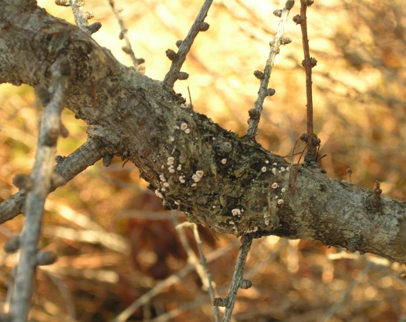
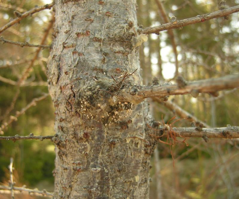
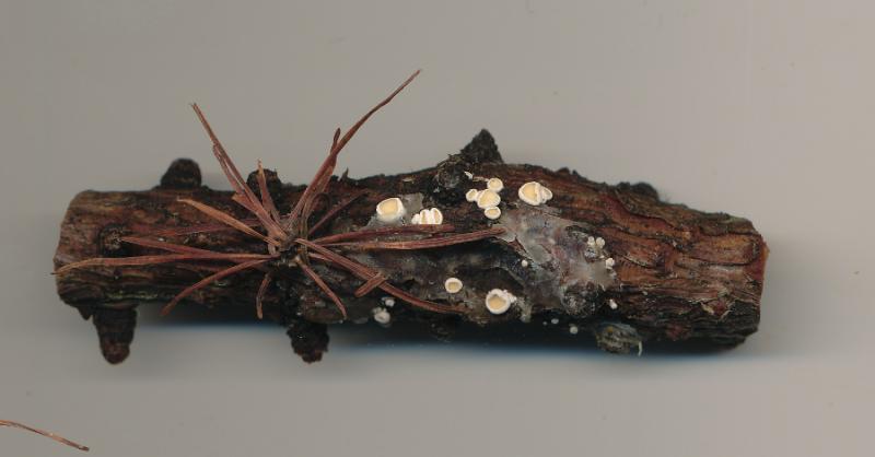
Where I live, near Saint john, New Brunswick, the fungus is very common and infects many or maybe all larches (Larix laricina). If you have that species you should be able to find swollen cankers on the branches, especially of young trees. The apothecia on the cankers are often accompanied by shiny layers of resin. The pictures are of this species as I find it near my home.
Dave
Didi you never see a red reactive apical ring in all these samples? Is there no occidentalis in your area?
See also our previous discussion
http://www.ascofrance.fr/search_forum/21908
There you gave the same ascus photo. Are these canker photos here also from the same specimen (Feb. 2013)?
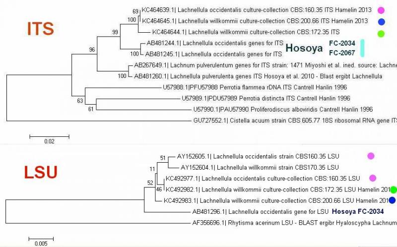
In the meantime further sequences have been deposited in GenBank.
I made a phylotree and again recognize the confusion around this complex.
Note that the very same CBS strain of occidentalis appears at two different positions (lilac), sequenced by Verkley and by Hamelin.
Zotto
The picture of asci was from twigs that had fallen from a tree during a winter storm. The cankers were photographed in April of the same year on different trees, so it is possible these are not both L. willkommii. I could test the material in KOH+Lugol now, but they may be too old. Perhaps I should collect some fresh material and try again. I believe both would be easy to find. If I find them, would someone like to sequence these? If so, cultures or apothecia?
Regards,
Dave
But Lugol directly without KOH should give a red reaction then.
Dave: The material can hardly be too old to test the iodine reaction. Only after 30-50 years this may happen that a red reaction changes to blue. So when you add Lugol directly you can see. But your ascus tips look too thin-walled to contain such an amyloid ring.
Pedro Crous from CBS, Netherlands, is interested in culturing and sequencing Helotiales, especially pathogens. Sending of air-dried apothecia should enable him to have active asci still. I can ask him.
Zotto
P.S. Just got his answer:
"Dave can send me the material or give it to Keith Seifert to bring in April, but I receive packages in good order from Canada - if it's a pathogen, I am hooked!"
Dave
Hi Zotto !
Question pour vous :
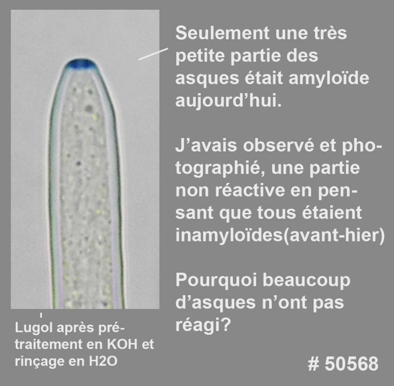
Pourquoi environ 90% des asques de l'échantillon sont inamyloïdes et que seulement une petite partie réagit ?
Le test a été refait, selon vos conseils, en mettant du Lugol après avoir prétraité dans le KOH, épongé et lavé avec H2O.
Seulement une petite partie des asques était amyloïde, les autres asques ne réagissant pas du tout, même après 1/2 heure.
Voici le résultat !
Roland