
16-04-2026 22:09
Buckwheat PeteHello, I'd like to ask about this older specimen:
14-04-2026 05:32
Ethan CrensonHi all, A few weeks back a friend pointed out som
12-04-2026 15:52
Gernot FriebesHi,I'm looking for help with this anamorph collect
15-04-2026 19:33
Fátima Durán ManzanequeHi!! I need help, I found this Ascomycete but I d
14-04-2026 21:52
Gernot FriebesHi,found on dead leaves of Carex elata. Conidia: 4
14-04-2026 20:31
Gernot FriebesHi,can this be Psilachnum lateritioalbum on Phragm
12-04-2026 17:56
 Hardware Tony
Found on dead stems in February earlier this year
12-04-2026 12:22
 William Slosse
In a dune grassland in Oostduinkerke (Belgium), on
11-04-2026 15:45
 Zuzana Sochorová (Egertová)
Please, could anyone send me this paper?Moyne G.,
11-04-2026 13:34
Artem PtukhaHello, I am seeking assistance with the identific
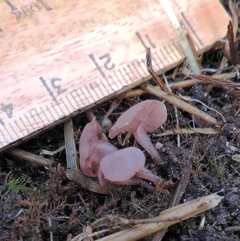
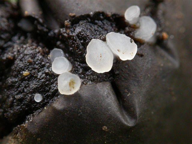
A colleague sent me an image a few of days ago of this fungus, growing on acidic soil in a damp area adjacent to boggy ground. The marker is in inches, but I estimate they are about 25-30 mm across.
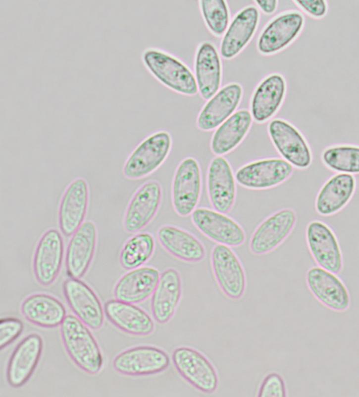
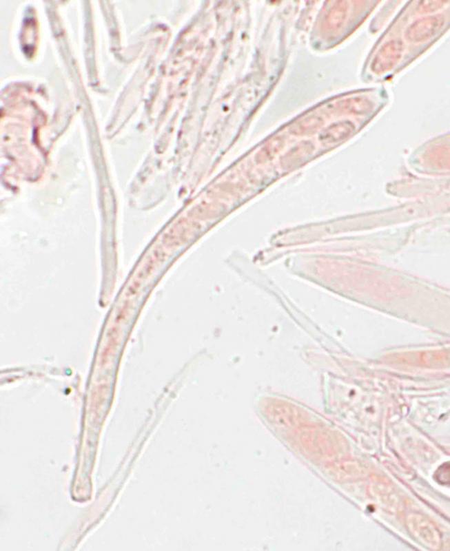
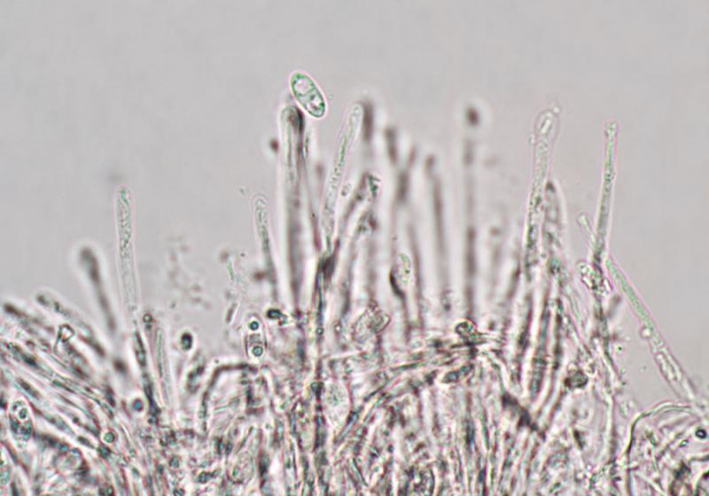
I subsequently received a tired specimen, but still fertile. The set specimen dried out too readily for a spore-drop but I have a decent image from a slide preparation with just the pressure from the coverslip, so should be more-or-less mature: 8.1-11.0 x 3.5-4.4 µm. A later sample had slightly large range: 9.9-12.1 x 3.8-4.4 µm.
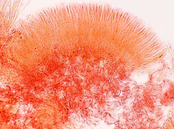
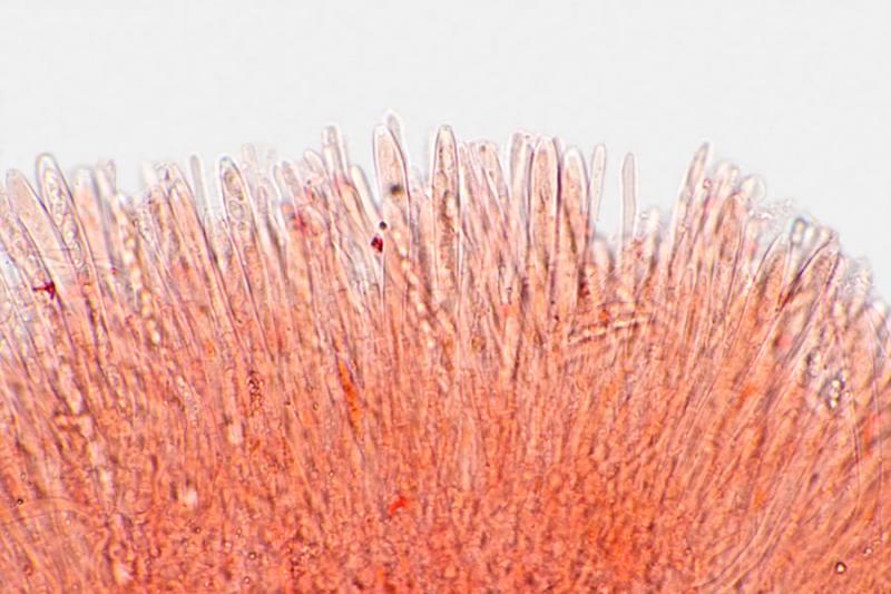
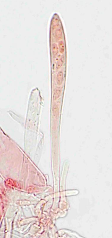
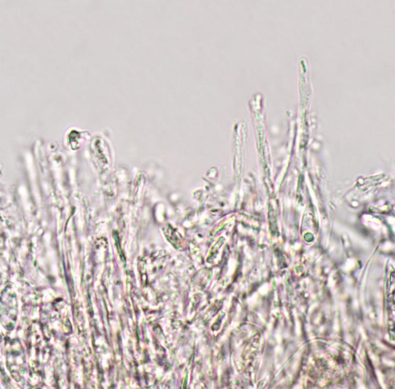
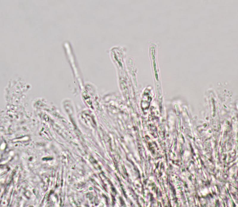
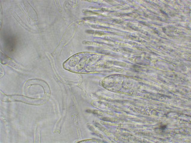
Asci: 90-103 x 8-9µm (small sample).
Paraphyses, heads: 3.0-3.3µm across.
Asci and paraphyses are of equal length.
The original thought was Ombrophila, but probably too small.
Possibly Sarcoleotia turficola, but the spores are too small.
Any ideas welcome.
Many thanks,
Chris

I agree with the genus Ombrophila. Is there any chance to know the substrate? I assume some decayed wood etc. buried in the soil?
Which country does the sample come from?
Zotto
It's in a friend's garden here on South Uist, extreme west of Scotland. There is scrub alder and willow close by, The basic substratum is peat but the area has had trees during the past 120 years - now gone.
I can inspect the site this week if necessary.
Chris
I do not see any crystals on your pics, which are typical of many species.
I compared Ombr. tetracladia as closest match, although I know it with shorter stalks and much much smaller (ca. 0.5-1 mm diam).
But with such large size, maybe it is a Roseodiscus? If there is no obvious gel in the medulla, I suggest comparison with R. formosus. This species was so far alwqys found in close association with Ceratodon purpureus (see Wieschollek et al. 2011, Z. Mykol.), though the spores are longer in this species, and its occurrence is in early spring.
Chris
Roseodiscus formosus does seem to have large spores and the paraphyses are curved. Also, it's not on the British list.
Ombrophila tetracladia isn't on the British list either and I can't find a complete reference to it. The spores are a good match but, as you say, the fruiting body is very small.
Chris

I have emailed you Zotto's Ombrophila key; while the teleomorph may not yet be listed as British, I encounter the anamorph Articulospora tetracladia very frequently when looking at Ingoldian fungi in stream-foam samples . . . .
amitiés
Chris
So when you still have the fungus fresh, please look in a water mount without applying pressure on the cover slip.
I attach an English translation of the Wiescholleck paper.
Zotto
 Wieschollek-etal-2011-Roseodiscus-Text-engl-0001.doc
Chris
I'll have another look at the fungus in water later today.
Chris
Best wishes,
Chris
Zotto
Chris
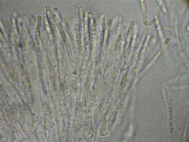
The VBs in the paraphyses are very well shown.
The substrate could be old fruits of apples? It is possible that these pics exist in
Ascofrance, but how to find them?
Hope that Michel can help.
Zotto
I found Rosoeodiscus formosus several times -it's certainly not the one at your disposal.
Cheers,
Marcel
Have read the avaiable data, I fully agree with you. Thanks for your thoughts.
Best wishes,
Chris

Hi Zotto,
Sorry I overlooked this post at the time it appeared on this site .
Yes of course those were my Coolpix micro pics with the beautiful rings -:)
Th ecollection was made in forêt de Mervent on 29 0 08 on old wild apples . (MH 40708) . I still have your pics you sent after receiving the collection , if necessary.
Amitiés
Michel

i overlooked this intersting thread. i would exlude r. formosus definitely, even in spore-shape. perhaps ombrophila rivulorum would be an idea, if we take the cristal-problem aside? how was the iodine-reaction? red or blue?
best
dirk
thanks! Now I have the problem that my computer is dead after 6 years of use, and I do not remember the folder where I had these pics when discovered last October. Ah, I found them, under O. tetracladia. But it is not the harddisk, it is the motherboard.
Zotto