
10-06-2026 21:16
 François Freléchoux
Bonsoir,Le dernier du jour, en attendant votre avi
11-06-2026 19:01
 William Slosse
Hello all,In an attempt to make a culture of a sus
11-06-2026 19:03
 Nicolas VAN VOOREN
Chers membres d'Ascofrance,Le site sera placé en

09-06-2026 18:32
Camille MertensSur morceau de roseau immergé 0,5 - 0,7 mm de dia
10-06-2026 12:54
Steve ClementsBonjour encore, Pouvez-vous m'aider, s'il vous pl
10-06-2026 21:07
François Freléchoux
Toutes les tiges de gentianes jaunes de l'an pass�
10-06-2026 13:41
François Freléchoux
Bonjour à nouveau, Voici une trouvaille d'hier.
10-06-2026 11:53
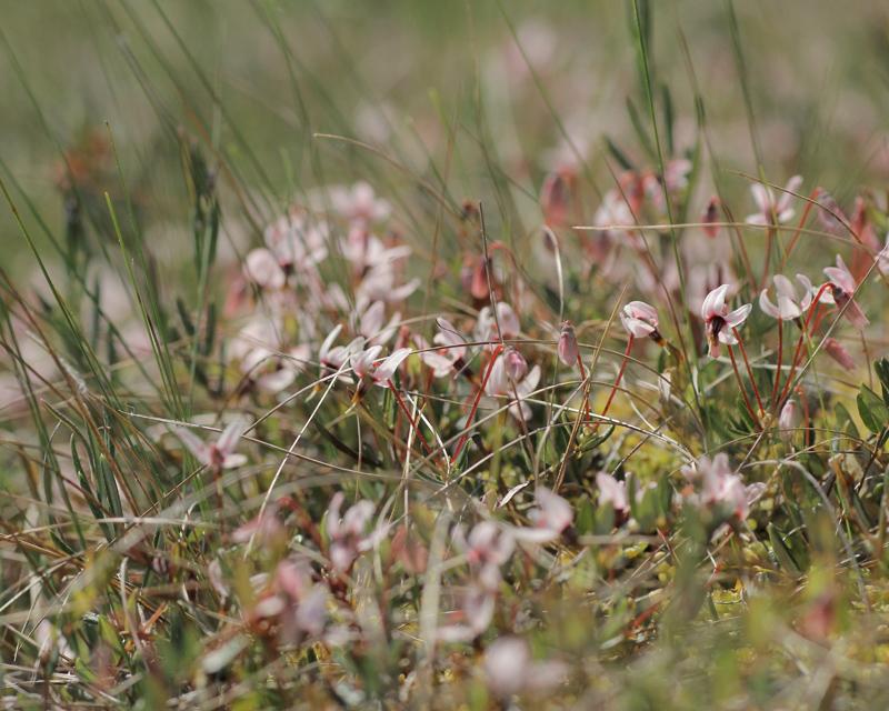
Steve ClementsBonjour, This disco is abundant on dead stems of
First and most easy searchable is Monilinia oxycocci, apothecia are arising from hidden inside of berries sclerotia just in early summer.
(#4168 - https://www.cubby.com/pl/%234168/_078e84b5ede740d4b778ee4cefc985a2)
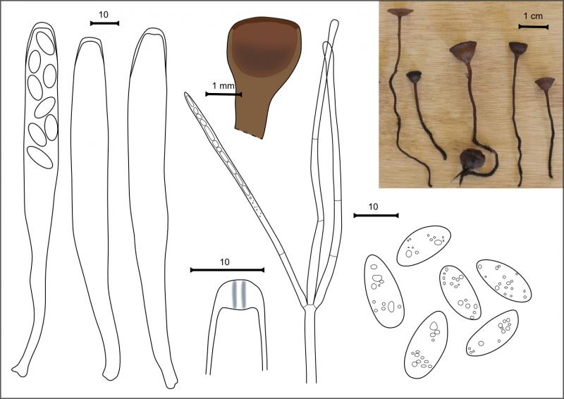
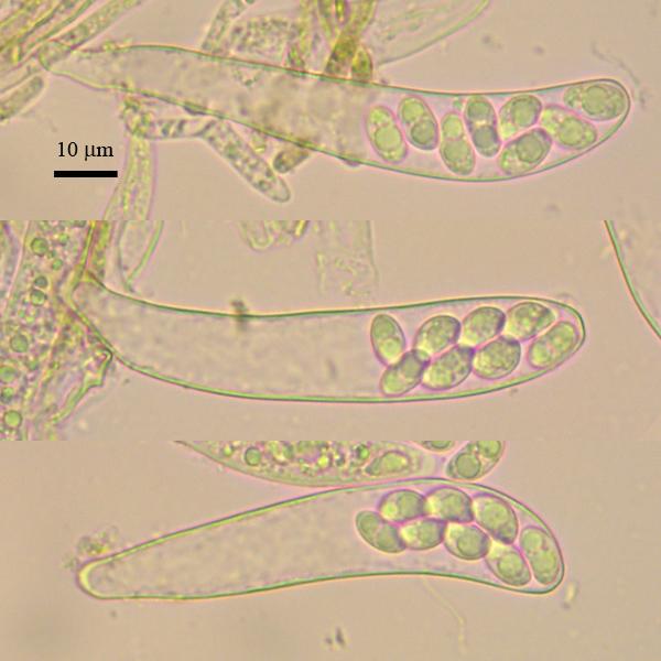
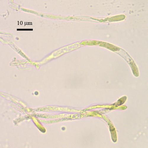
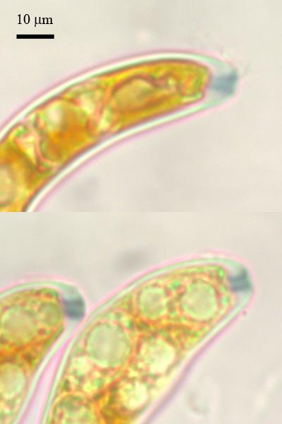
Another discomycetes is very tiny and spotted only under the lens. It have bold greenish discs, asci with amyloid ring, and fusoid curved spores. Tips of paraphyses and cells of excipulum are filled with greenish oil guttules, which determine the apos color.
(#4167 - https://www.cubby.com/pl/%234167/_b3e70f926f3743f8a78f23259118e517)
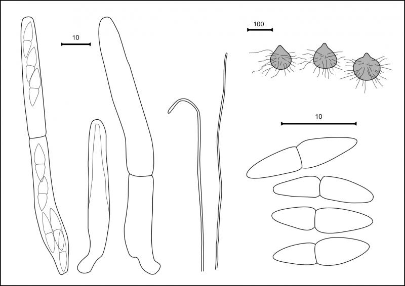
Small hairy perithecia was spotted at one old berry, and it is a new species in my bog collection. Have no idea of genus and species yet. Fissitunicate asci, presence of paraphyses, two-celled hyaline spores and hairy perithecia should assign to a certain one.
(#4170 - https://www.cubby.com/pl/%234170/_06ba95dd77ae4a9a8d473890c016b729)
Physalospora cf. vaccinii is quite common, but most perithecia are underdeveloped now.
(#4165 - https://www.cubby.com/pl/%234165/_48b3da7f0f454a4a87be8106a37db0a1)
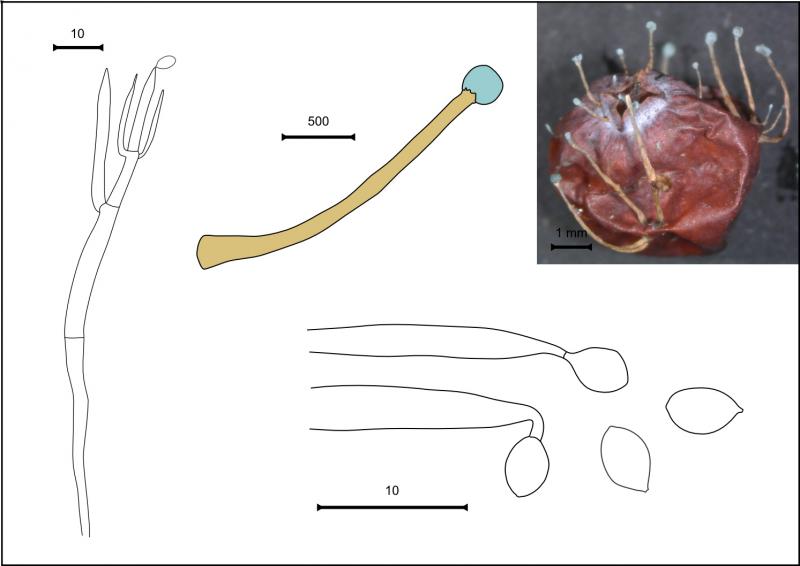
Now the anamorphs are coming. Probably some of them are connected with the species mentioned above, but they will wait for later identification. The first forming large sinnemata in the shape of matches with long stalks and blue heads. They are common at old berries (strange love of acidic fruits:). Quite beautiful, bright, easy distinguished, but not identified yet.
(#4173 - https://www.cubby.com/pl/%234173/_e603d10334a744b782938657c9840d82)
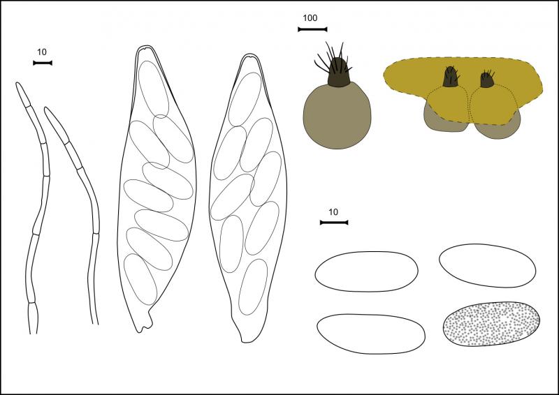
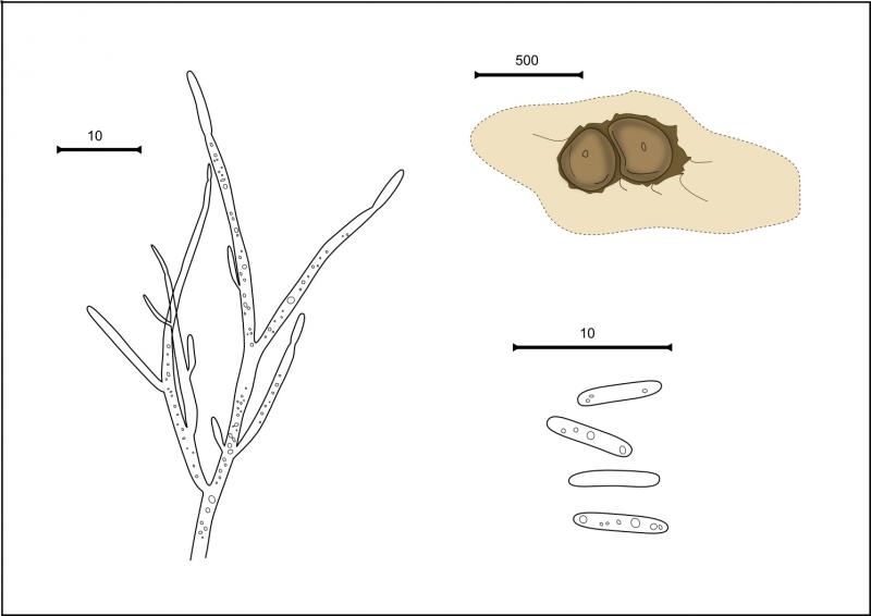
Next imperfect species is forming sporodochia: disc- to cushion-shaped dark brown bodies. Richly branched conidiophores crowded inside, forming rod-shaped conidia.
(#4169 - https://www.cubby.com/pl/%234169/_508653b1601f4e7a8db51b2b8fc4884d)
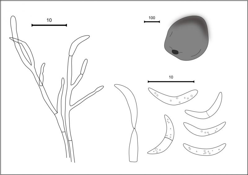
And yet another anamorphic species was with picnidial conidioma. The black spherical bodies with small raised operculum filled inside with branched conidophores with lunate hyaline conidia.
(#4174 - https://www.cubby.com/pl/%234174/_b4fa81dc9da14246be87e080cca7fc07)
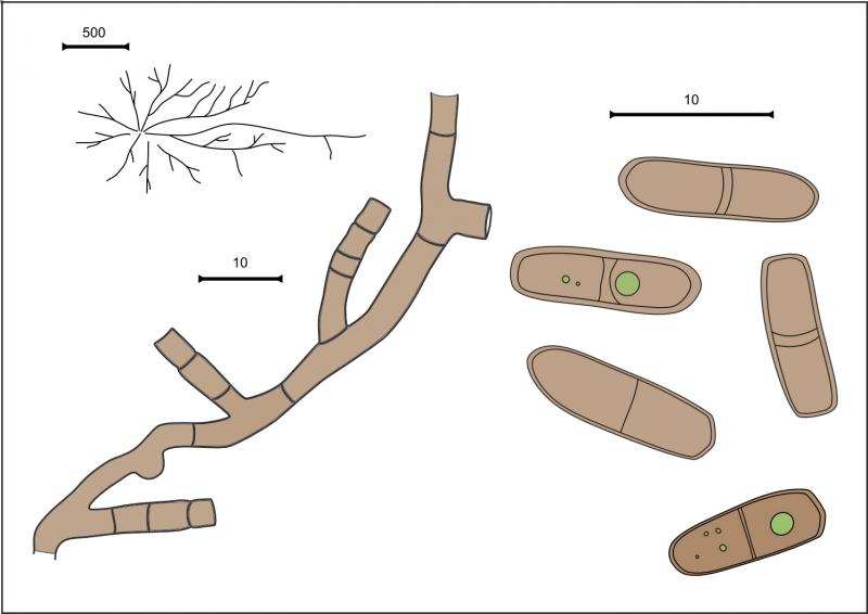
Some hyphomycete forms small dark appressed colonia at the upper leaf surface, and under microscope brown hyphae with brown two-celled conidia, born inside conidiogenous cells are seen.
(#4175 - https://www.cubby.com/pl/%234175/_0b95ce9bd1204777a279dc80e59c9cca)
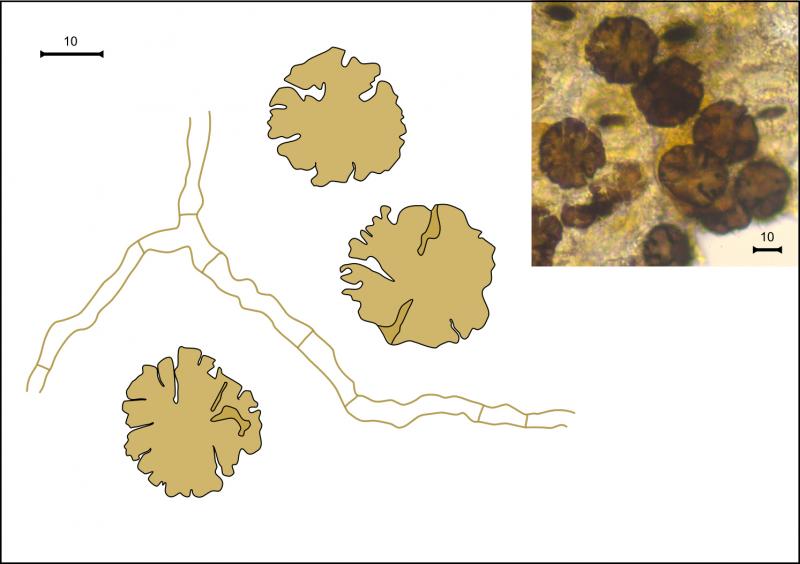
The last one constantly met is somehow doubtful, probably it is not anamorph but undeveloped stage of some species. It composed from round lobed rosettes, brown and thick, surrounded by brown hyphae.
(#4172 - https://www.cubby.com/pl/%234172/_c552074929b74b63b80e94fe7aeb5ac0)?

the second species could be a Calycellina. Did it grow on the stems? Did you study also the excipulum? The greenish contents are vacuoles, not oleaginous. I wonder whether the spore surface turns lilac in aqueous Cresyl Blue, as many Calycellinas do.
Your statements on the photos ("vital in water") is often not true, many images show dead asci. Also the images are too many, and therefore time-consuming to scroll.
Zotto

Good luck Walter
I will consider a Calycellina when will be working with literatue; it was on leaves; excipulum was studied, (a link below), its cells also has greenish vacuoles (thanks for correction) and the edge made with the same obtuse cells (without any hairy structures).
You are right, Zotto, i will add later the compilative photos and there will be a few of them showing necessary informaiton. All photos are only row collection, it is also usefull for measurements, but difficult to watch. I will improve this when will reach my linked computer (with better internet there).
A "vital" code word - i use it for specimens examined in vital stage (compared to studied from rehydrated specimens). Probably, asci may be dead since the frbs has been exposed to the sun in situ, or other reasons. I don't know ).
Penicillium vulpinum - thanks for this, i will study its description later.
Nina.?
The dead state is rather obvious on your photos. Due to the high number of photos it is difficult to point to a single one. You have both living and dead asci, and they look very different. When the VBs are greenish and refractive, the paraphyses are alive. It can also be a result of preparation. It is a frequent mistake to consider everything vital what is from fresh specimens.
Zotto
At this photo it seems that outer surface, but not sure. Yes, at the base cells isodiametric (about 5-8 mk) and becoming longer, hyphoid (cells 10-20 mk long, 4 mk broad).
https://ws.cubby.com/p/_b3e70f926f3743f8a78f23259118e517/%234167/%234167-excipulum-vital-water-x400-0001.jpg/436360633
Nina
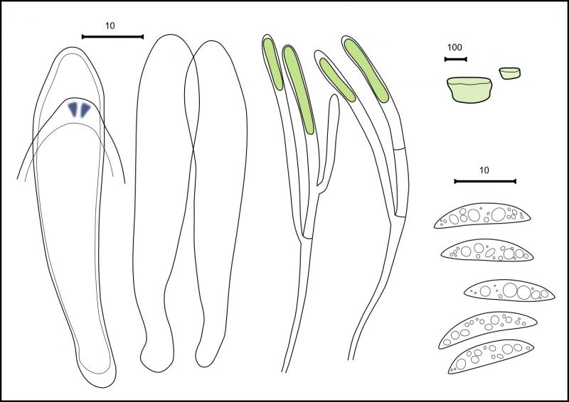
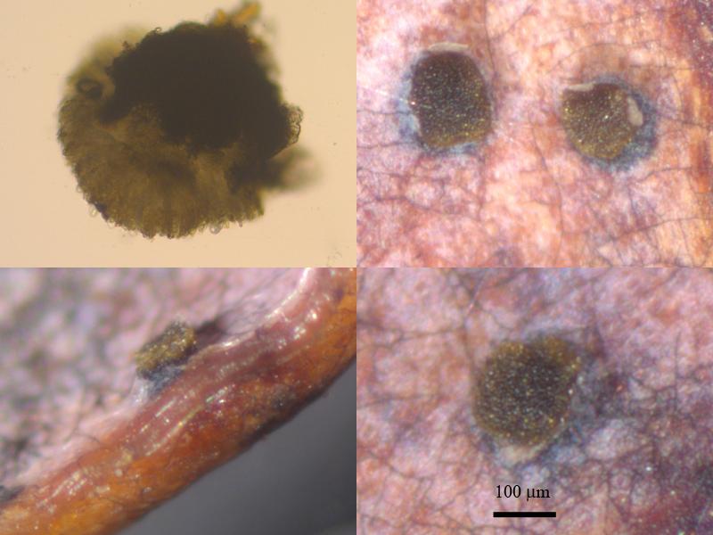
Searching cranberry litter today, i have found similar (Calycellina?), but another exemplar. Also rare species, single collection though hundreds of leaves were looked out. It has similar VBs filled paraphyses and cells of excipulum, but the spores are different.
Its description and photos:
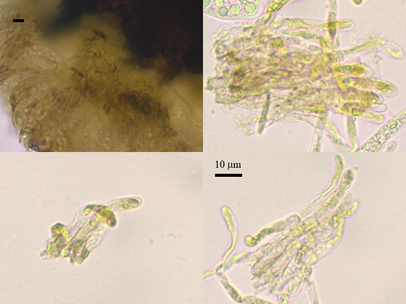
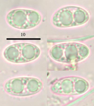
Apothecia pustulate, greenish, bursting through epidermis (base deepened in leaf tissue), 150–220 mk in diameter, 50 mk thick; several apos nearby at one leaf.
Excipulum very scarce, at base from irregular brown cells, and at upper part from prismatic, end hyphoid cells to 25 long and 3 thick, filled with greenish vacuoles; asci clavate, clampless, with euamyloid ring, about 82 x 15; paraphyses cylindrical, with obtuse tips, filled with greenish content (dead in studied specimen since drying in situ), about 73 x 3.7; spores ellipsoid, slightly heteropolar, with many round oils, 10.7 (10–11.8) x 5.7 (5.3–6) (n=12).?
No idea what on Ericaceae might exist in Trochila. Possibly undescribed. Nothing in my database.
Zotto
The apothecia were dryed at substrate already, and that is why the paraphyses are dead, i think. I will look further to encounter more collections of this species.?
Nina.
https://svampe.databasen.org/observations/10358814