12-06-2026 14:50
 François Freléchoux
Bonjour, Voici la brève description d'une Mollis
10-06-2026 21:16
François Freléchoux
Bonsoir,Le dernier du jour, en attendant votre avi
11-06-2026 19:01
 William Slosse
Hello all,In an attempt to make a culture of a sus
11-06-2026 19:03
 Nicolas VAN VOOREN
Chers membres d'Ascofrance,Le site sera placé en

09-06-2026 18:32
Camille MertensSur morceau de roseau immergé 0,5 - 0,7 mm de dia
10-06-2026 12:54
Steve ClementsBonjour encore, Pouvez-vous m'aider, s'il vous pl
10-06-2026 21:07
François Freléchoux
Toutes les tiges de gentianes jaunes de l'an pass�
10-06-2026 13:41
François Freléchoux
Bonjour à nouveau, Voici une trouvaille d'hier.
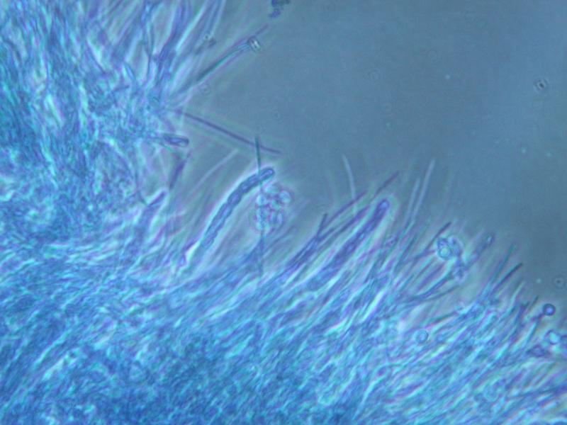
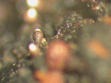
It occurs on exudate of wounds of Prunus pensylvanica in PEI and NB Canada.
The ascomata are about 200-400 um tall and 150-200 um wide, and urceolate in shape. The walls are light brown and become melanized as it matures. Internally, the walls consist of linear, periclinally arranged hyphae. The asci are narrowly cylindric (c. 35 x 4 um), and lack an evident apical apparatus. They break down at maturity, producing a dry mass of spores that collects around the ostiole, and within the cavity in the upper part of the ascoma. Externally, the spore mass appears white to very pale yellow. The ascospores are uniseriately arranged, 8 per ascus, colourless, unornamented, and c. 3-4 x 2-3 um. They are ellipsoidal, but slightly compressed on the long axis.