
10-06-2026 21:16
 François Freléchoux
Bonsoir,Le dernier du jour, en attendant votre avi
11-06-2026 19:01
 William Slosse
Hello all,In an attempt to make a culture of a sus
11-06-2026 19:03
 Nicolas VAN VOOREN
Chers membres d'Ascofrance,Le site sera placé en

09-06-2026 18:32
Camille MertensSur morceau de roseau immergé 0,5 - 0,7 mm de dia
10-06-2026 12:54
Steve ClementsBonjour encore, Pouvez-vous m'aider, s'il vous pl
10-06-2026 21:07
François Freléchoux
Toutes les tiges de gentianes jaunes de l'an pass�
10-06-2026 13:41
François Freléchoux
Bonjour à nouveau, Voici une trouvaille d'hier.
10-06-2026 11:53
Steve ClementsBonjour, This disco is abundant on dead stems of
Bonjour.
Merci de venir à mon secour pour détermine ce que je pense être un asco,
Récolter sur Astragallus glycyphyllos.
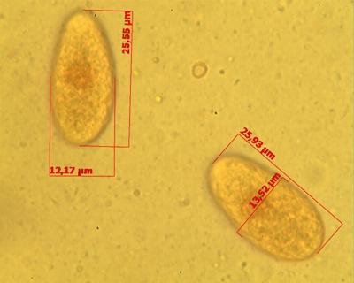
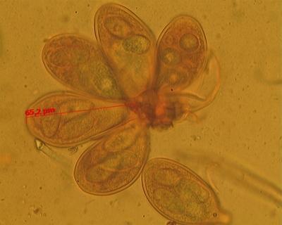
Les asques (avec 4 spores) mesurée (63.58-65.2) X 34.81 µm.
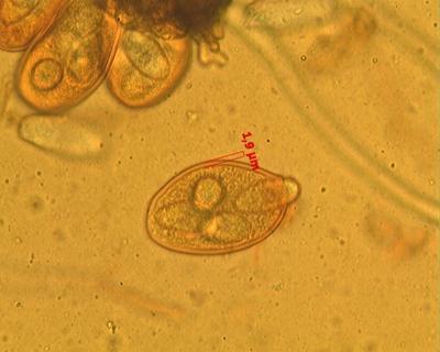
Les spores épaisses de 1.9 µm. mesurée (25.55-25.93) X (12.7-13.52) µm.
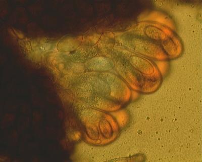
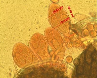

As for me your fungus asci and ascospores are similar with Erysiphaceae representative.
What can you say about the fruit bodies and appendages of fruit bodies?
Alex
Quant à moi vos champignons et asques des ascospores sont similaires avec le représentant Erysiphaceae.
Que pouvez-vous dire sur les corps des fruits et des appendices des organes de fruits?
Alex

C'est un oïdium, donc bel et bien un asco: une Erysiphale.
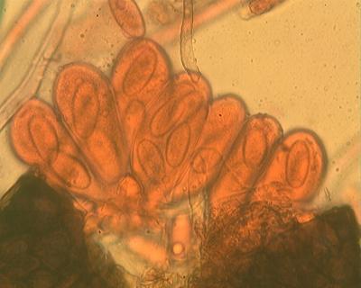
Sur une photo, on voit les fulcres dirigés vers le haut (ce sont les poils des petites cléistothèces). Cela correspond bien avec Microsphaera astragali (maintenant Erysiphe astragali) qui pousse sur Astragalus glycyphyllos. Les dimensions des spores et les asques correspondent bien aussi. Je l'ai eu au Mont des Pins.
Regarde dans la doc que je t'avais envoyée sur les oïdiums, notamment les publications de Léon Slupinsky: il est dedans.
Amitiés - LUC.
(bon, maintenant je termine de préparer mes affaires: je pars en Champagne pour quatre jours, faire de la myco notamment, et je reviendrai sans doute avec des ascos.)
Description from Braun, U. 1987, Nova Hedwigia, Beih. 89: 298
Erysiphe astragali DC., Fl. Fr. VI, p. 105 (1815).
= Microsphaera astragali (DC.) Trev.
Alphitomorpha holosericea Wallr., Verh. Ges. naturf. Freunde BerIin 1, p. 41 (1819) , type host - Astragalus glycyphyllos. A. astragali (DC.) WaIIr., Ann. Wetter. Ges. N. F. 4, p. 244 (1819). E. holosericea (Wallr.) Link, in L., Sp. Pl. 4, 6(1), p. 115 (1824). Alphitomorpha sericea Wallr., Fl. Crypt. Germ. 2, p. 757 (1833), type host - Astragalus glycyphyllos. Microsphaera holosericea (Wallr.) Lév., Ann. Sci. Nat., bot., 3. sér. ,15, p. 159 & 381 (1851). Trichocladia astragali (DC.) Neger, Flora 88, p. 351(1901).
IIl.: Léveillé (1851, pl. 9, fig. 27); Tulasne & Tulasne (1861, pl. 5, fig. 2); Magnus (1899, pl. IX, fig. 5-7); Salmon (1900, pl. 3, fig. 47-51); Blumer (1933,p. 335, fig. 133; 1967, p. 245, fig. 81); Vasjagina et al. (1961, p. 279, fig. 91); Sandu-Ville (1967, p. 286, fig. 50); Moro?kovs'kij et al. (1969,p. 74, fig. 35); Zhao (1979, p. 94, fig. 52); Yu & Lai (1981,p. 35, fig. 2); Braun (1984c, p. 240, fig. 40); Salata (1985, pl. IX).
Lit.: Saccardo (1882,p. 12); Salmon (1900,p. 127); Jaczewski (1927,p. 300); Blumer (1933,p. 334; 1967, p. 245); Vasjagina et al. (1961, p. 278); JuneIl (1967,p. 54); Sandu-ViIIe (1967, p. 258); Moro?kovs'kij et al. (1969,p. 73); Eliade (1976,p. 194); Bunkina (1979,p. 87); Zhao (1979,p. 93); Yu & Lai (1981,p. 35); Salata (1985,p. 147).
Exs.: F. exs. suec. 1190. Fl. Exs. Austr.-Hung. 3576. Fuck. , F. rhen. 694. Krieger, F. sax. 1222. Rabenh. , F. eur. 439, 2413. Rabenh., Herb. myc. 469. Rehm, Ascomyc. 448. Romell, F. exs. scand. 62. Roum. , F. gaIl. exs. 1164, 3141. Sacc. , Myc. ven. 148. Syd. ,Myc. germ. 2520. Syd. , Myc. march. 979. Thüm. , F. austr. 459. West., Herb. Crypt. Belg. 1059.
Mycelium amphigenous, effused or irregular patches, persistent or subpersistent, conidia formed singly, ellipsoid-cylindric, ca 30-45 x 16-24 µm (fresh), appressoria lobed, conidiophores erect, foot-cells cylindric, 30-50 x 8-10.5 µm, followed by two shorter cells, sometimes by one longer cell. Cleistothecia scattered or subgregarious, 80-155 µm in diam, cells irregularly polygonal, obscure, ca 6-20 µm diam, appendages equatorially arising or somewhat from the upper half of the ascocarp, ca 2-12 times as long as the cleistothecial diam, ca 5-25, basal part mostly distinctly curved, appendages horizontally spread or usually turning towards one direction, sometimes fasciculate, flexuous, hyaline or coloured near the base, aseptate or with 1-2(-3) septa near the base, thin-walled to moderately thickwalled, usually thin-walled above and thick-walled towards the base, smooth to rough, 5-12 µm wide, apex mostly simple, some appendages dichotomously branched, 1-3 times, branchings loose and wide, lax, tips straight or recurved, primary branches sometimes recurved, occasionally forked in the lower half, asci 5-14, mostly shortly stalked, 50-85 x 25-50µm, ascospores (2-)3-5(-6), ellipsoid-ovoid (-subglobose), (15-)18-26x 10-16 µm. Pl. 79.
Lectotypus: on Astragalus glycyphyllos L., France, "Erysiphe astragali, No. 235," herb. de Candolle (G).
Hosts and distr.: on numerous host species of the genus Astragalus, Aragallus and Oxytropis, Fabaceae; all Europe, Mediterranean region, Asia Minor to Central Asia, Siberia, China, Far East of the USSR, Japan, Pakistan, North America (USA).
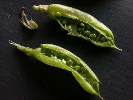
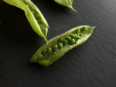
Les fruit en forme de petit haricot qui font entre 35 / 40 mm de long épais de 4 mm avec a l'intérieur comme des petit pois de 1 mm dans une double enveloppe et ils sont relier a la nervure centrale par un petit filamant.
Joseph
J'ai été intéressé par les organes de fruits de champignons, pas des Astragakus :-) C'est un petit boules de couleur qui ont été obtenues avez indiqué dans la photo ascospores.
Alex