
28-05-2016 11:13
 Castillo Joseba
agrupados y meegrosAlguna sugerencia ?Josebaa
28-05-2016 23:10
 Bernard CLESSE
Voilà une Peziza trouvée à 10 mètres de chez m
27-05-2016 22:39
Bernard CLESSE
Bonsoir à tous,Une récolte de cet après-midi qu

26-05-2016 11:33
Pascal RIBOLLETBonjour Forum,Je suis aux prises avec un pyréno r
27-05-2016 17:21
Bernard CLESSE
Bonjour à tous,Trouvé cet après-midi cet asco s
27-05-2016 17:26
Bernard CLESSE
Dans le même site (aulnaie marécageuse avec ruis

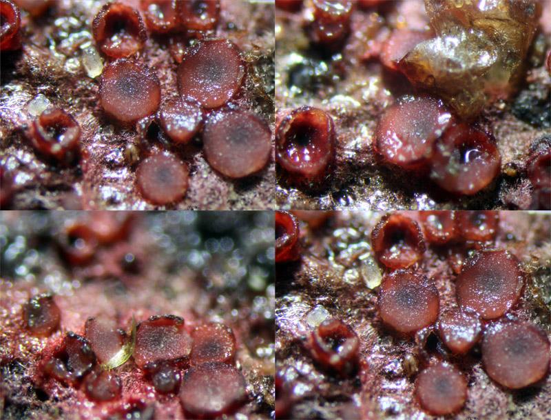
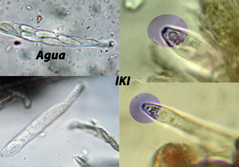
Roja
Castillo Joseba,
28-05-2016 11:10
en madera de Fagus o CprilusAlguna sugerencia?
Joseba


Adam Polhorský,
28-05-2016 22:11
Re : Roja
Hi Joseba,
this is Patinellaria sanguinea, the red subiculum is very typical.
Cheers,
Adam
this is Patinellaria sanguinea, the red subiculum is very typical.
Cheers,
Adam
Castillo Joseba,
29-05-2016 21:36
Re : Roja
Gracias Adam