
26-04-2023 18:11
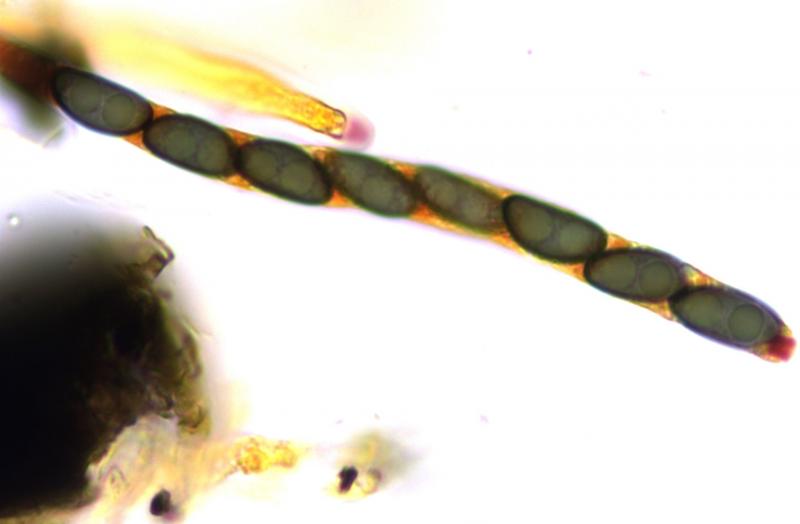
Joaquin MartinHiI found this pyreno on fagus wood.As you can see
28-04-2023 08:57
Zuidland PeterHello all,I have what I think is a Hydropisphaera
28-04-2023 16:47
 Bernard Declercq
Dear Forum,Following was collected today:Conidioma
27-04-2023 06:49
Zuidland PeterHello all,I found a few of these small apothecia (


24-04-2023 23:39
Ismael WindThis species was growing on oenothera. Spores are
25-04-2023 14:13
Rob van KruiningHello,I found this white Propolis in Norway on a b
25-04-2023 15:56
Juuso ÄikäsThese little black pyrenos were growing yesterday
30-03-2013 21:25
 François Valade
HelloA small yellow Bryoscyphus sp? on moss, Pleur
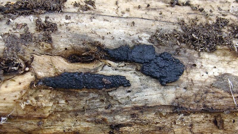
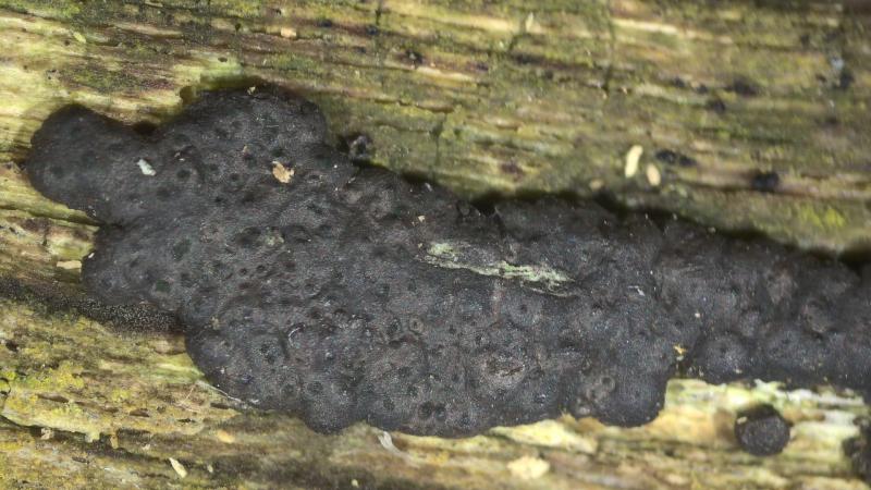
Nemania serpens - colliculosa
Joaquin Martin,
26-04-2023 18:11
I found this pyreno on fagus wood.
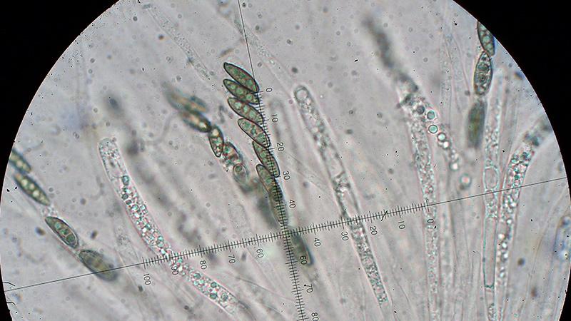
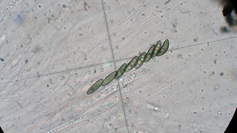
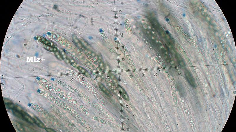
As you can see it has a blue apical reaction with Meltzer, which would lead me to N.colliculosa.
the sporal measurements are:
(12.3) 13.3 - 15.4 (16.7) × (3.7) 3.9 - 4.8 (5.1) µm
Q = (2.6) 2.8 - 3.7 (4.1) ; N = 40
Me = 14.4 × 4.4 µm ; Qe = 3.3
My questions are: is this Nemania colliculosa ? N.serpens var colliculosa ? or N.serpens
Thank you, best regard
Thomas Flammer,
28-04-2023 13:33
Re : Nemania serpens - colliculosa
I was yesterd confronted with Nemania serpens var. serpens.
I observed that serpens has a red, not a blue reaction with Barals.
Spores 11.1 - 14.3 x 4.6 - 5.8 ?m
Joaquin Martin,
30-04-2023 10:31
Re : Nemania serpens - colliculosa
Thanks Thomas.
I will leave it as N.colliculosa, with doubts.
Mycobank has it legitimated as a species.
Best regards
I will leave it as N.colliculosa, with doubts.
Mycobank has it legitimated as a species.
Best regards