
16-08-2024 14:16
 B Shelbourne
I found the (asco)stroma of an Epichloe sp. a few

05-08-2024 08:26
 Lothar Krieglsteiner
French Alps, 13.7.24, on twig of either Alnus alno
09-08-2024 08:22
Andreas LoosliHello Everybody, my Name is Andyi am new here in
11-08-2024 10:28
Henri KoskinenOrange apothecia 2-8 mm, on sandy roadside soil am
13-08-2024 17:20
Lothar Krieglsteiner
.. a collection from 19.3.2008, Hessen, Germany, o
04-08-2024 19:30
Lothar Krieglsteiner
French Alps, on Corylus, together with Orbilia tra
22-04-2024 20:38
 Miguel Ángel Ribes
Good afternoon.Does anyone know this anamorph?It g
12-08-2024 09:28
Miguel Ángel Ribes
Good morning This small (about 1 mm wide), stalke
Epichloe sp with hyperparasite
B Shelbourne,
16-08-2024 14:16
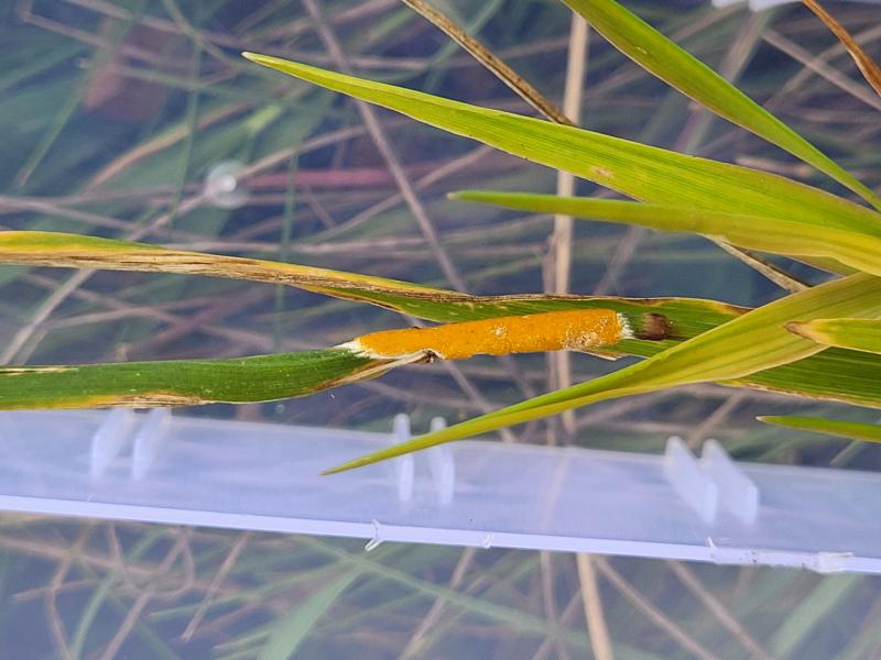
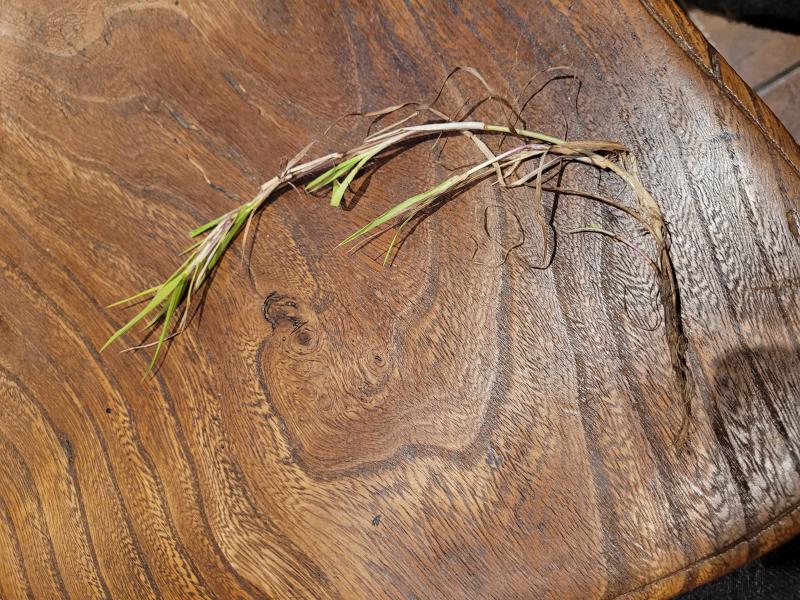
I found the (asco)stroma of an Epichloe sp. a few days ago in southern England, and there are also signs of a green-spored hyperparasite that could be Bionectria epichloe.It has been very interesting to learn about the genus Epichloe, and I would appreciate any feedback on identifying the species.
I can't confidently identify the host grass, but my best guess is genus Poa and then E. typhina (s.l.), the latter may fit the macro, and asci and spore sizes. Although I did find some shorter spores that I thought could be part spores, but I'm not convinced now. Next time, I think capturing ejected spores would be helpful.
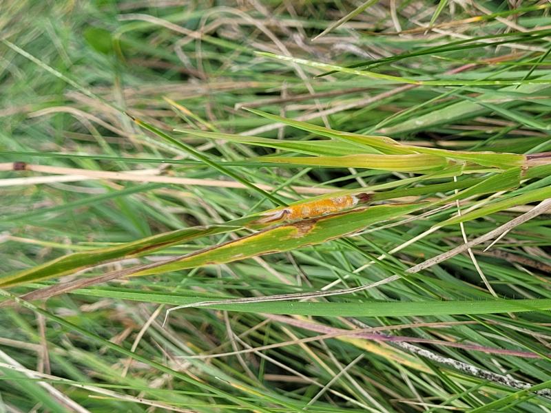
The habitat is an open chalk hill (downland) with several grasses and small plants around, 128m altitude, at a junction of paths, surrounded by semi-natural grasslands, and agricultural, horse, and hay fields, in the South Downs.
*Host
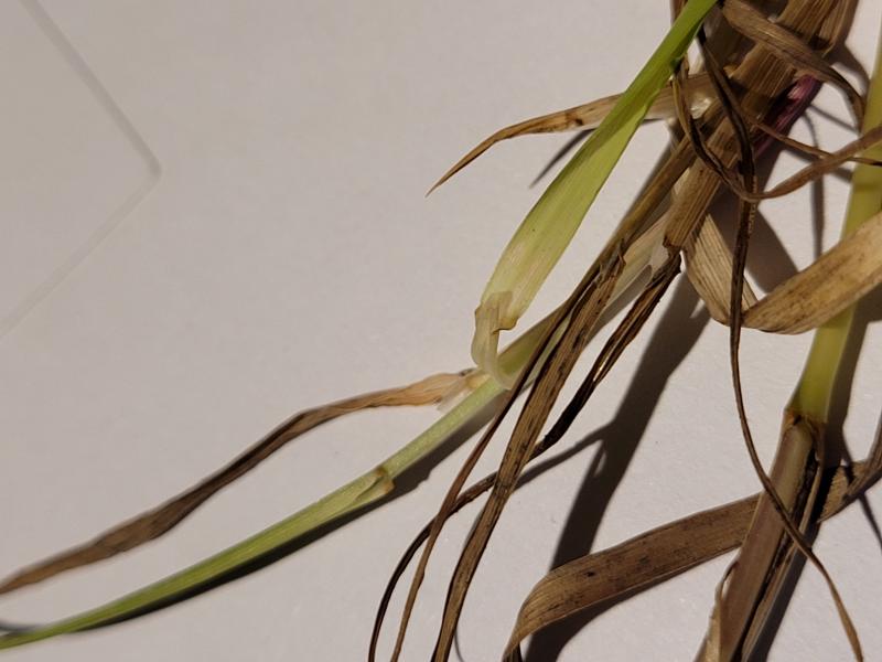
I have no experience with vegetative characters of grasses, and I thought it may be Agrostis stolonifera, but after more reading and looking at the plant then my best guess is Poa cf. pratensis.
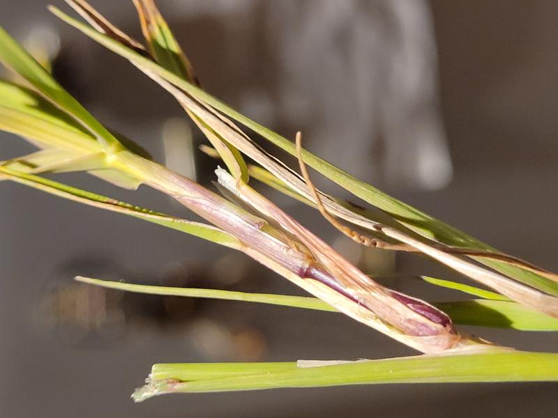
A low grass, with bluish, tapering leaves, shiny on the underside, folded in the stem, the sheaths have a rough texture (especially on the main stem), many have turned purple with age, the ligules are white, membranous, broadly acute, several measured 1-4 mm, no apicules identified, all parts appear hairless, the base of the main stem is almost white with some old brown sheaths, the lateral roots are small and there are no rhizomes.
*Epichloe
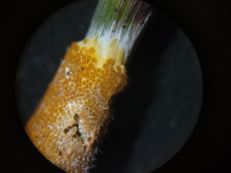
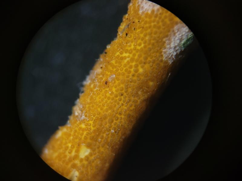
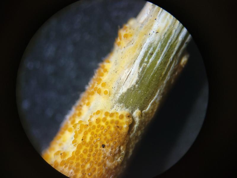
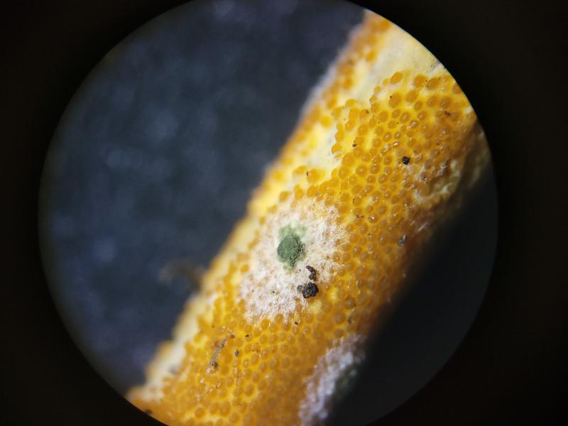
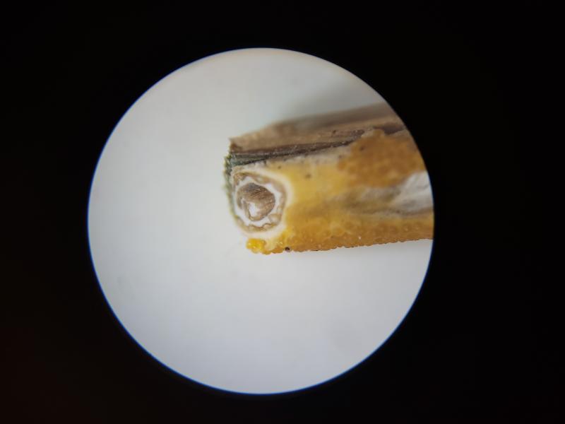
Macro: Stroma covering most of an internode and part of the flag leaf on the living main stem of a grass, ~20 x 2-3 mm, signs of necrosis in the top part and further development of the stem and inflorescence is arrested. Underneath is a dense white mycelium with a smooth, spongy appearance, on the outside yellowish-organish with immersed perithecia (fertilised), ~0.2-0.3 mm diameter, approximately several thousand, gelatinous texture and appearance, densely arranged, orange, globose-pyriform shape, ellipsoid from above.
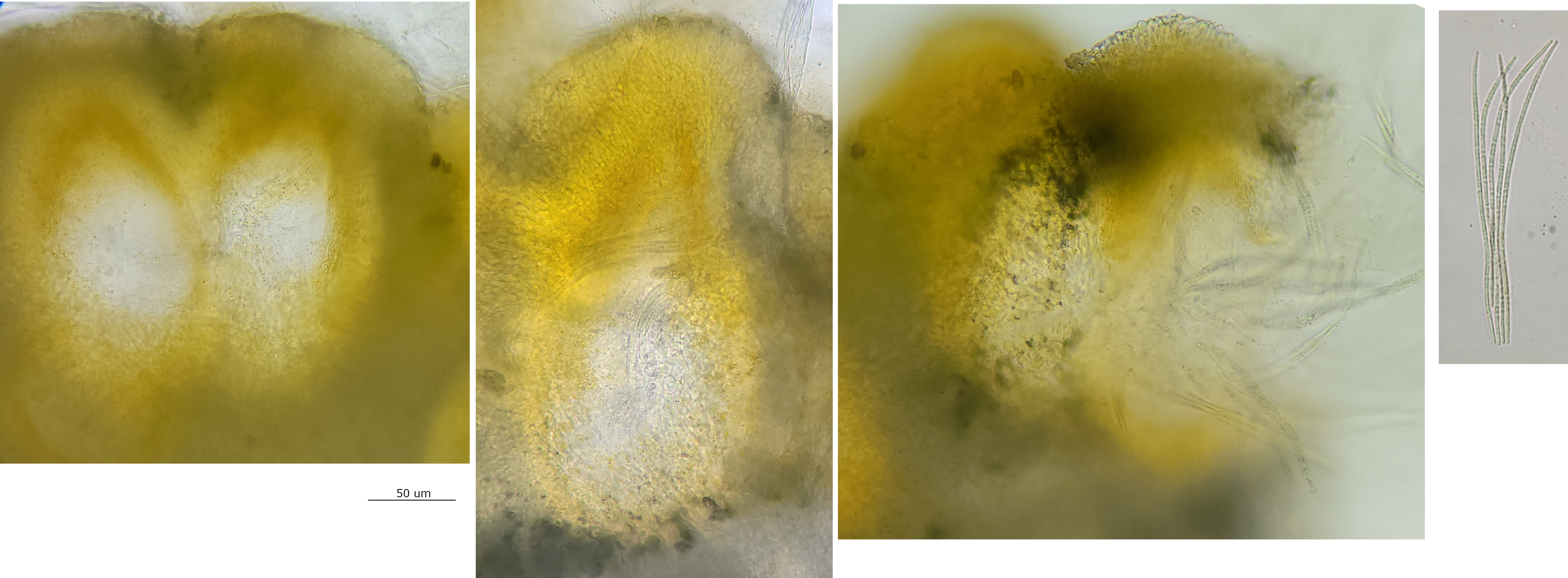
Perithecia with pyriform shape, neck extending towards the surface, neck protruding from stroma, inner margin and neck strong pigmented, with many long and slender asci developing from the base, no paraphyses (as expected), many small yellowish globose vacuoles in surrounding hyphae.
Asci narrow-cylindrical, sometimes appearing to bulge in the middle, ~150-180 x 8-10 um, shorter asci possibly immature, and probably slight underestimate for mature asci, apex hemispherical, highly refractive, inamyloid, poroid, apparently dark unrefractive cap developing in maturity, appears easily separated from ascus, bases with croziers.
Spores: Hyaline, multiseptated, with many small to tiny greenish vacuoles, usual reaction from contents when cells die, two shapes apparently associated
- filiform, ~(110) 140-170 (180) x 1-2 um, several counted with 6-9 septa,
- some ~45-80 um long, apparently less septa but hard for me to see.
Not confident that these are part spores, but not sure if immature or what the explanation might be.
In the lower layers textura globosa with small yellowish vacuoles, possibly the stroma.
*Hyperparasite
Macro comparable to Bionectria epichloe, no micro currently.
On the surface of the stroma are patches of white mycelium, some appearing to have green conidia developing at the centre (e.g. photo 9), some signs of the mycelium invading the stroma, and also suppression and necrosis (e.g. photo 6).
 40x-0001.jpeg
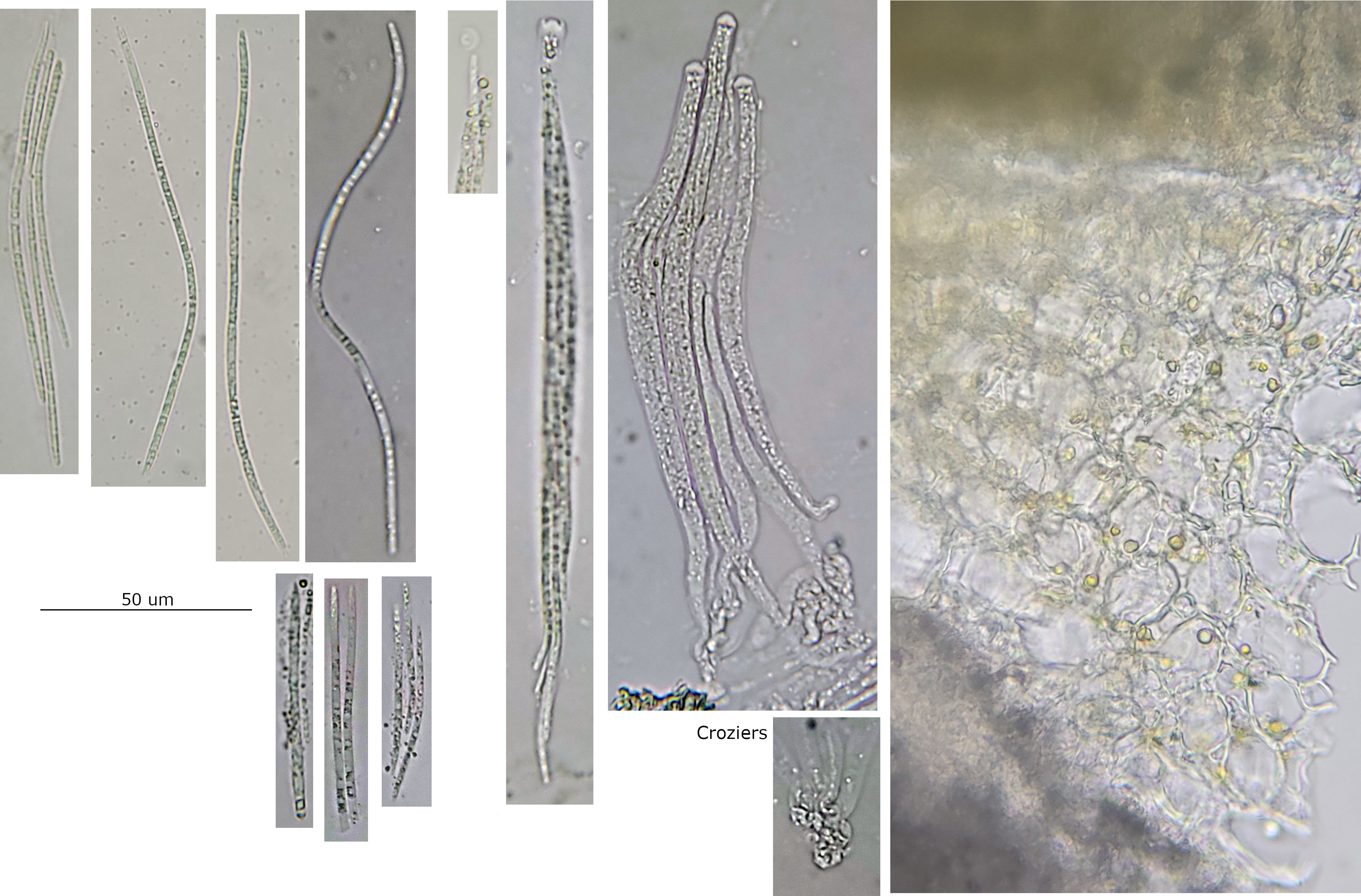 60x-0001.jpeg