03-09-2016 23:47
 Rubén Martínez-Gil
Hola a todos. Parece que últimamente Bernard y y
01-09-2016 19:23
 Andgelo Mombert
Bonjour à tous,Mon premier message sur ce forum,
02-09-2016 15:23
SYLVAIN ARDBonjour, Je viens proposer mes services bénévol


30-08-2016 17:38
Dragiša SavicHi, one question,I found today Lasiosphaeria hirs
22-04-2016 09:23
 Castillo Joseba
Buscando una especie, estoy viendo otrasEsta me
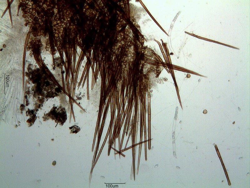
Hola a todos.
Parece que últimamente Bernard y yo hemos estado en la misma zona ya que hemos encontrado cosas muy similares...
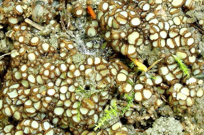
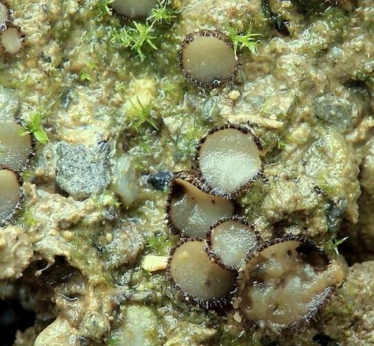
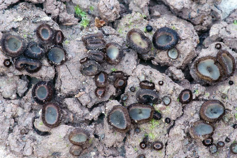
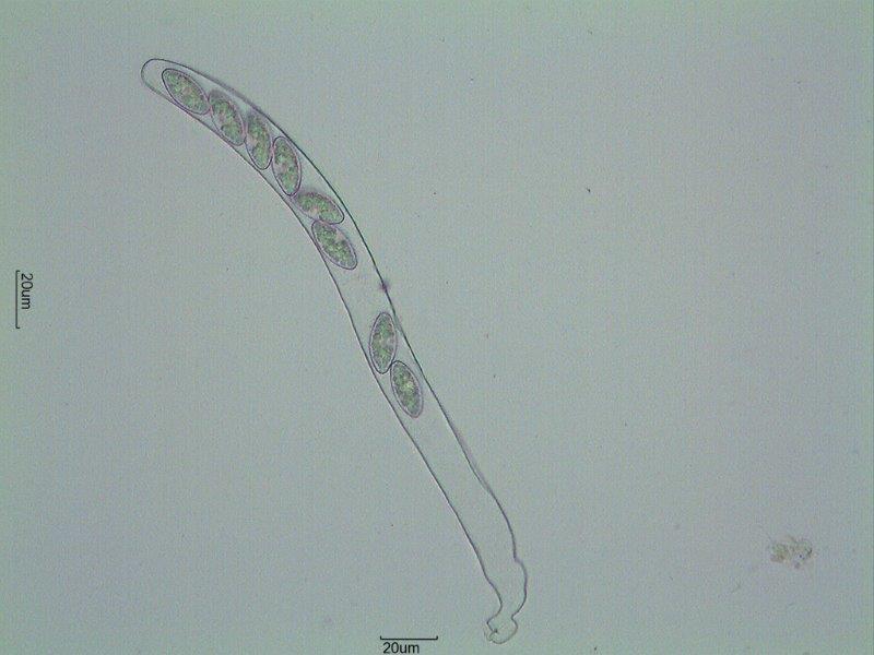
Subo unas fotos de un asco que encontré sobre tierra en zona calcárea.
Miden hasta 5 mm de diámetro.
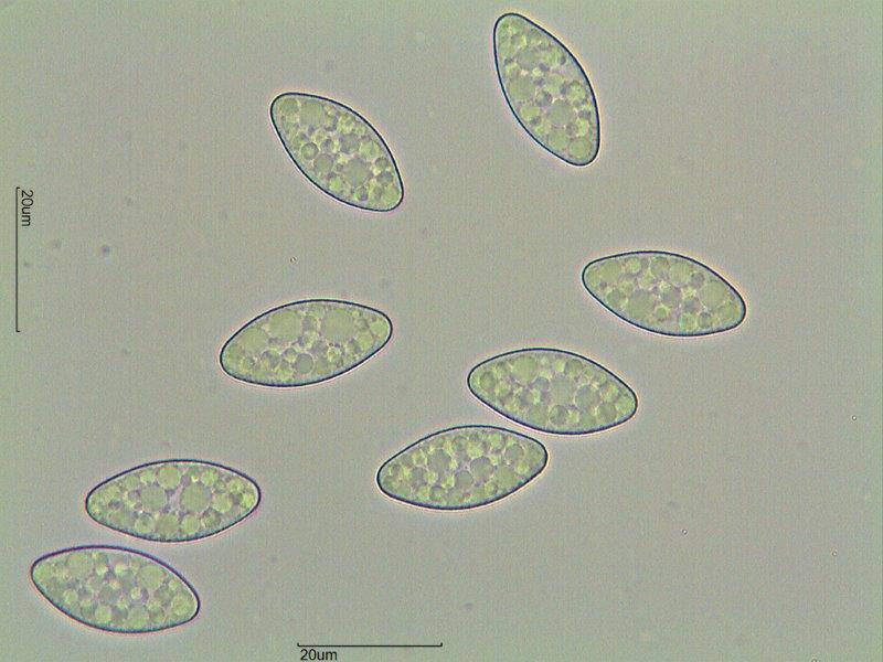
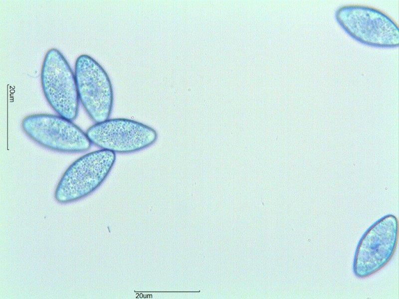
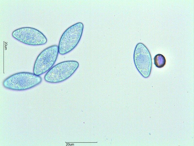
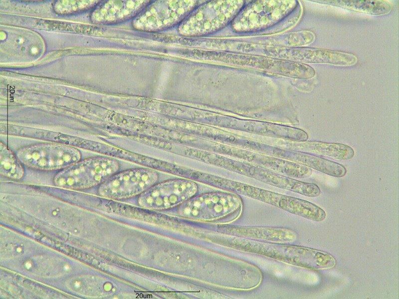
Esporas con verrugas finas, de 21-25 x 9,5-11 micras.
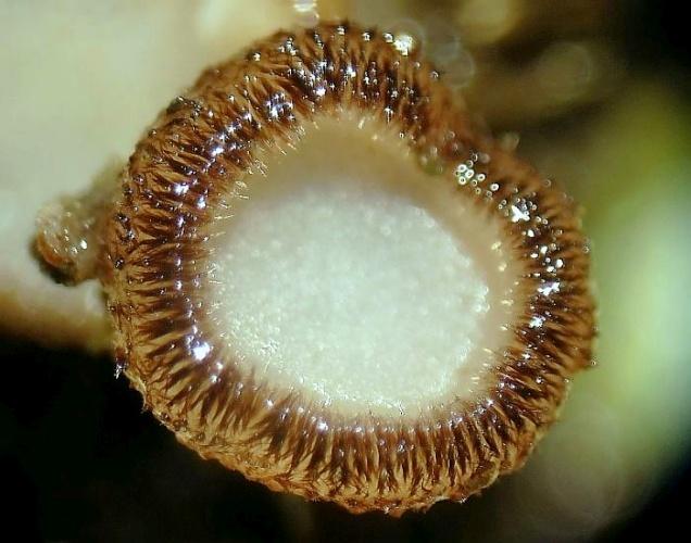
Ascas pleurorrincas, operculadas.
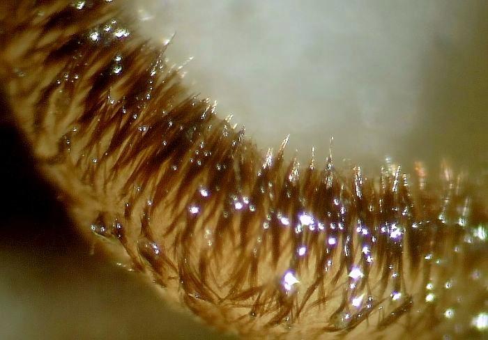
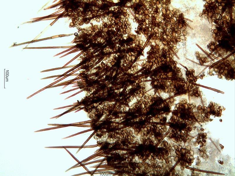
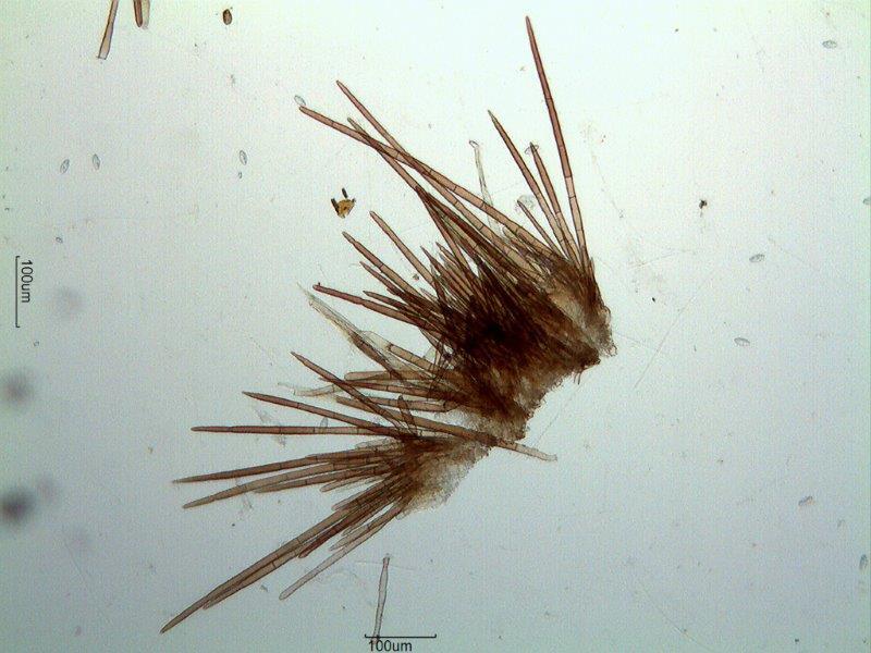
Pelos hasta unas 480 micras.
Yo pensé en Trichophaea gregaria, ¿Está sinonimizada con Trichophaea hybrida? ¿Son diferentes?
Gracias por su ayuda.
Rubén

Tr. hybrida is a synonym, see
VAN VOOREN, NICOLAS (2014): Contribution à la connaissance des Pézizales (Ascomycota) de Rhône- Alpes– 2re partie. Cahiers de la FMBDS nr. 4.
too.
Greetings Peter.