
20-07-2017 14:19
Watt JohnIl y a trois jours, j'ai trouve cet asco qui resse
20-07-2017 13:40
 Ralf Dahlheuser
I found this type for the first time and I want to

04-07-2014 11:33
Marja Pennaneneven on their dead leaves.Hi there,these beauties
16-07-2017 00:24
Thorben HülsewigHi there,today i was very suprised, because i foun
18-07-2017 19:59
 Andgelo Mombert
Bonsoir,Un Trichopezizella sur Epilobium.Spores 10
17-07-2017 18:35
Andgelo Mombert
Bonjour à tous,Sur bois mort de Alnus albobetula,
17-07-2017 20:12
 Castillo Joseba
De ayer en madera de fagusA ver que os pareceJoseb
Hypocreopsis????
Elsa Sousa,
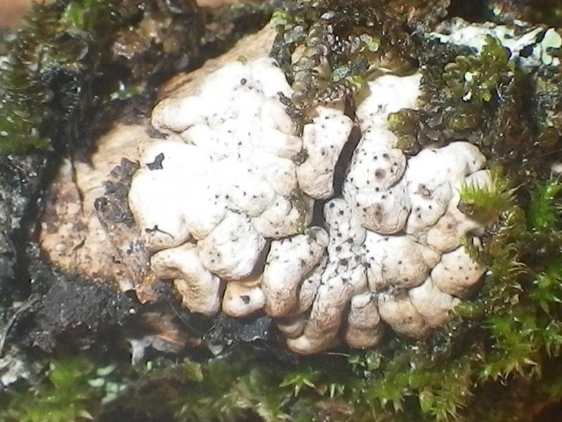
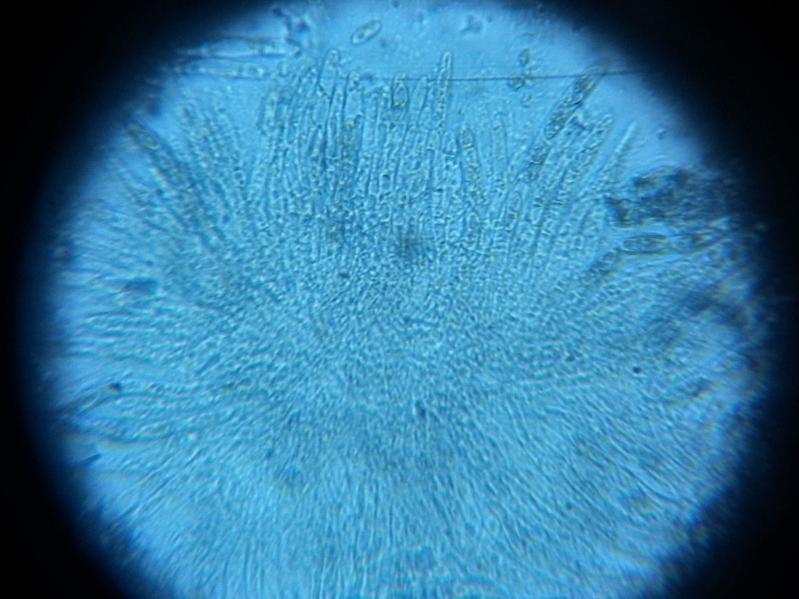
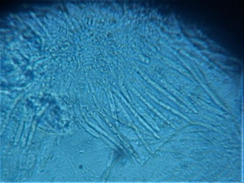
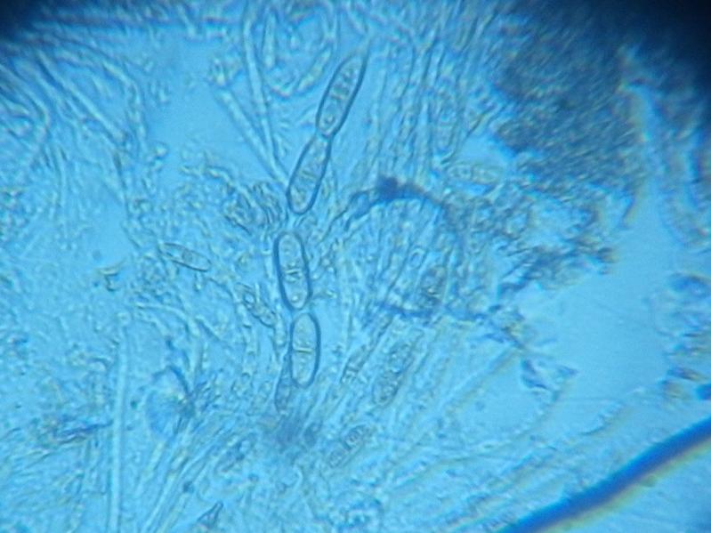
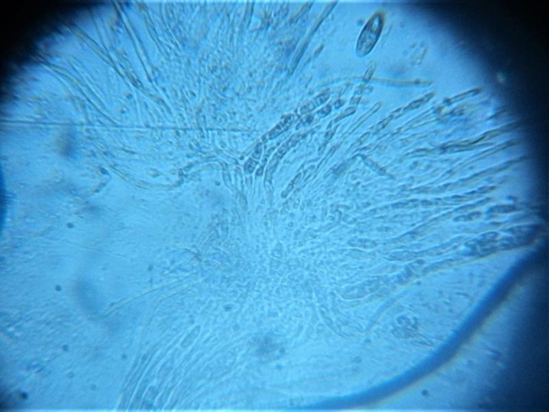
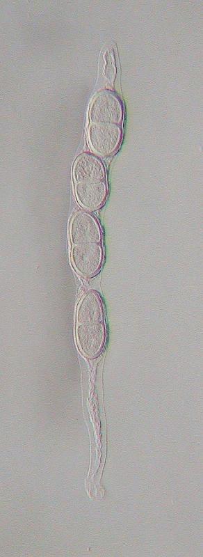
18-07-2017 23:40
Good evening,
I found it on a stick of unknown tree.
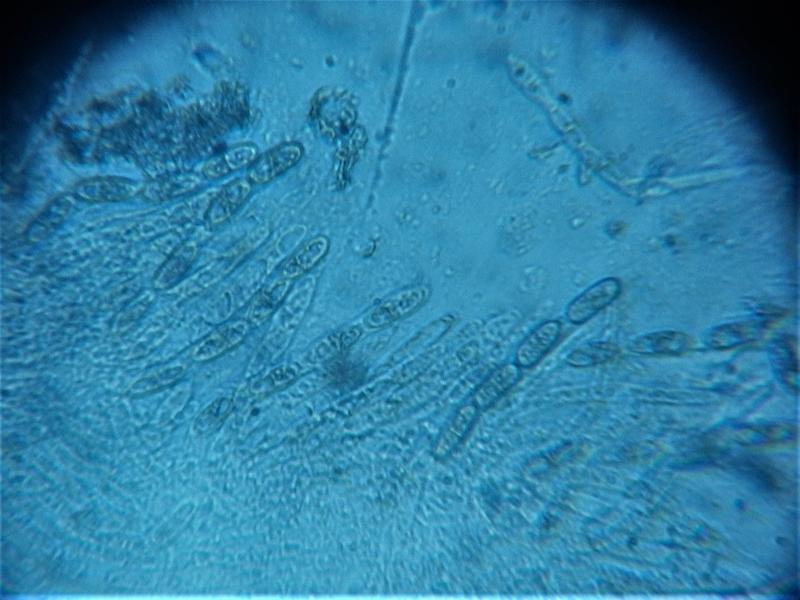
Asci 4-spored
(30.1) 31.3 - 34.6 (34.8) × (10.9) 11.1 - 12.9 (13.9) µm
Q = (2.4) 2.41 - 2.87 (2.9) ; N = 7
Me = 33.1 × 12.2 µm ; Qe = 2.7
I would appreciate any help.
Thank you very much.
Elsa Sousa
Christian Lechat,
19-07-2017 04:56

Re : Hypocreopsis????
Dear Elsa,
your fungus is a new species of Hypocreopsis, I am working with the same and it would be great if you could send me your collection. I attach two images of my specimen.
Thank you very much in advance:
My address:
Ascofrance
Christian Lechat
64 route de Chizé
79360 Villiers-en-Bois
FRANCE
your fungus is a new species of Hypocreopsis, I am working with the same and it would be great if you could send me your collection. I attach two images of my specimen.
Thank you very much in advance:
My address:
Ascofrance
Christian Lechat
64 route de Chizé
79360 Villiers-en-Bois
FRANCE
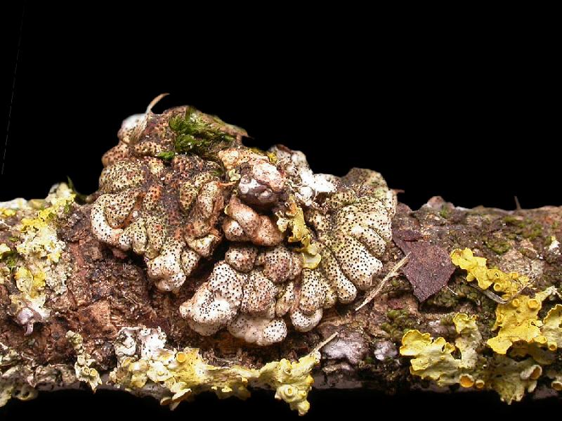
Thomas Læssøe,
19-07-2017 13:56
Re : Hypocreopsis????
Dear both
what is the host of this new taxon? (Hymenochaetopsis?). And its current known distribution?
cheers
Thomas
what is the host of this new taxon? (Hymenochaetopsis?). And its current known distribution?
cheers
Thomas
Elsa Sousa,
19-07-2017 14:06
Re : Hypocreopsis????
Dear Thomas,
I'm not sure about the host of my specimen, once I took it from the ground on a small park. However I can take photos of the different trees and the tree nearby the stick. I am sure that there are oaks and some other ornamental trees.
Dear Christian, I'll send you the fungus, but now it's not attached to the stick, is there any problem? Do you want the piece of the stick too?
Regards,
Elsa
Christian Lechat,
19-07-2017 15:47
Re : Hypocreopsis????
Hi Thomas,
my specimen is on Peniophora sp. on Syringa in France .
Regards
Christian
my specimen is on Peniophora sp. on Syringa in France .
Regards
Christian
Elsa Sousa,
19-07-2017 21:00
Re : Hypocreopsis????
Interesting Christian... my stick has a peniophora too, so I'll send both the slice and the piece of stick. My bet is that the stick is from an Acer Negundo (It was the closer tree).
Regards
Regards
Christian Lechat,
20-07-2017 16:01
Re : Hypocreopsis????
Ok Elsa,
I'm waiting for your sending.
All the best,
Christian
I'm waiting for your sending.
All the best,
Christian