
08-12-2013 12:58
 Rubén Martínez-Gil
Hola a todos.Pongo unas fotos de una Otidea que en
11-12-2013 08:37
Ibai Olariaga IbargurenHi there, I am trying to identify a non-strom
08-12-2013 22:17
Daniel GhyselinckGood evening,On dead stem of Urtica, with Leptosph
09-12-2013 01:09
Rubén Martínez-Gil
Hola a todos.Pongo unas fotos de Spathularia que e
09-12-2013 12:43
 Christian Lechat
Dear friends,I would need of the following papers:
06-12-2013 19:18
 Joop van der Lee
Found on horse dung, Fruitbody: 87.36x61.38 um; c
04-12-2013 13:30
Garcia SusanaHello I found this ascomycete on sheep dung.Apoth
06-12-2013 10:51
GÜNGÖR HalilHello ?s there anybody who can help me about this
05-12-2013 11:58
 Vasileios Kaounas
Found 05-12-13, Attica Greece, in soil, in forest
05-12-2013 23:25
 Maren Kamke
Hi everybody,I need your help with this asco. Apot
Otidea especie
Rubén Martínez-Gil,
08-12-2013 12:58
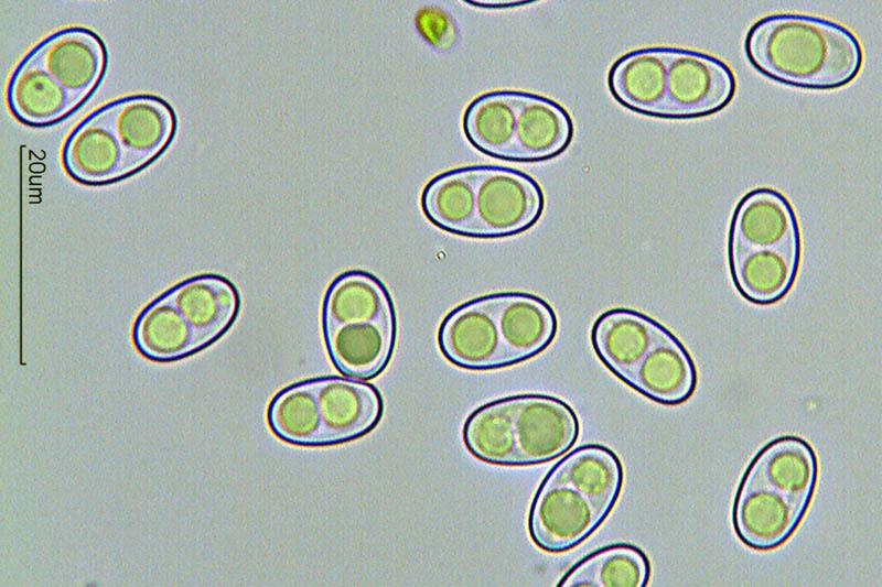
Hola a todos.Pongo unas fotos de una Otidea que encontramos hace un par de días en un pinar de P. nigra con boj y algún quejigo suelto.
Miden hasta 4,5 cm de largura.
Estaban casi enterradas entre la pinocha y sólo se les veía la parte superior (+- 1 cm).
Al apartar la pinocha, encontré más ejemplares de menos de 1 cm de diámetro y que al principio me parecieron Peziza de color algo más oscuro.
Medida esporal de 10-12 x 6-7 micras.
No he apreciado tonos rosas en himenio y por ello me decidí a mirarla.
Al secarse, el himenio se ha quedado de color naranja y el exterior de color beige claro. El margen se ha quedado de color pardo.
Yo pensé en O. onotica, pero sin tonos rosas. ¿Podría ser O. formicarum?
Gracias a todos.
Rubén


Ibai Olariaga Ibarguren,
10-12-2013 23:25
Re : Otidea especie
Hola Rubén,
Creo que se trata de Otidea subformicarum, una especie muy próxima a O. formicarum que va a ser publicada pronto. Se distingue de O. formicarum por tener esporas un poco más largas (9.5–11 × 6–7 µm en formicarum). La localidad tipo de O. subformicarum está en el valle de Pineta (Huesca).
Saludos,
Ibai.
____________
Dear Ruben,
I think it is O. subformicarum, a species closely related to O. subformicarum that will soon be described. It differs from O. formicarum in the slightly larger spores (9.5–11 × 6–7 µm en O. formicarum). The type locality of O. subformicarum is in Pineta (Huesca, Pyrenees).
Cheers,
Ibai.
Rubén Martínez-Gil,
11-12-2013 12:32
Re : Otidea especie
Gracias por la información, Ibai.
También, creo que O. formicarum es algo más oscura??
Un saludo
Rubén
También, creo que O. formicarum es algo más oscura??
Un saludo
Rubén