
12-06-2026 14:50
 François Freléchoux
Bonjour, Voici la brève description d'une Mollis
10-06-2026 21:16
François Freléchoux
Bonsoir,Le dernier du jour, en attendant votre avi
11-06-2026 19:01
 William Slosse
Hello all,In an attempt to make a culture of a sus
11-06-2026 19:03
 Nicolas VAN VOOREN
Chers membres d'Ascofrance,Le site sera placé en

09-06-2026 18:32
Camille MertensSur morceau de roseau immergé 0,5 - 0,7 mm de dia
10-06-2026 12:54
Steve ClementsBonjour encore, Pouvez-vous m'aider, s'il vous pl
10-06-2026 21:07
François Freléchoux
Toutes les tiges de gentianes jaunes de l'an pass�
10-06-2026 13:41
François Freléchoux
Bonjour à nouveau, Voici une trouvaille d'hier.
 I thought these tiny whitish ascomata look like a Hyaloscyphaceae sp., but it seems to be a difficult and broad group. I have no experience with the distinguishing characters and couldn't find any recent key to the genera. Any feedback appreciated.
There are some similarities to Hyaloscypha minuta, but the spore size (in particular) seems to be aberrant. I have more material, but the size makes it quite difficult to manipulate the ascomata and it would help to know which features to focus on.
The hairs look quite short or scarce (not noticeable under stereoscope) and plain, but the paraphyses seem Hyaloscypha-like (filiform without VBs). I couldn't match the features well with any species in Huhtinen's monograph on Hyaloscyphus (croziers, hemiamyloid rings, spore size), and I also considered Hyphodiscus (Hyphodiscaceae), but the disc isn't smooth, and the hairs don't seem right.
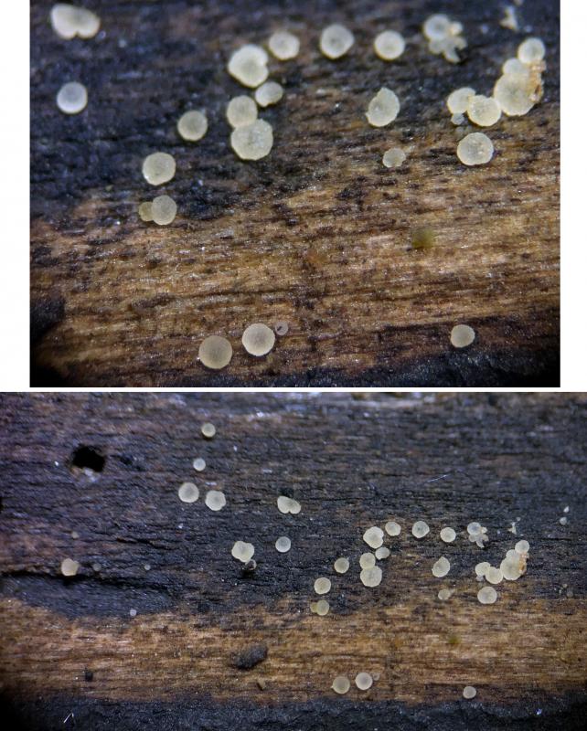
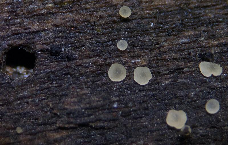
Habitat: Observed in January, on decaying (unidentified) deciduous wood, decorticated and often blackened parts, small pile of logs, presumably hygric, shady area, mixed deciduous woodland, southern England.
Associates: Several types of conidia found around base, many ascomata arising from or close to dematiaceous spore mat, on the same pile of logs - Calycina cf. citrina (previously), Lachnum impudicum, Orbilia eucalypti (previously), Trichoderma strictipile (aged ascomata), Mollisia sp., Peniophora sp.
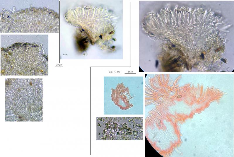
Preparation: Stored for a day in a damp container attached to wood, slides prepared from two ascomata, sections attempted but only some success with second ascomata, progressively squashed, mounted in tap water or KOH with IKI or CR applied afterwards.
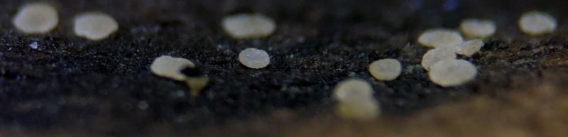
Ascomata: Hyaloscypha minuta-like, gregarious to clustered, superficial, < 0.5 mm diameter (maybe even smaller max), whitish-ochraceous, possibly more yellow with age, translucent, pruinose, some with lumps of orange exudate, initially cupulate to turbinate, then discoid to scutellate-pulvinate, often appearing sessile and appressed in maturity, no clear differentiation between receptacle and disc, occasionally uneven margin in maturity.
Reactions:
- IKI (~1%): Apical rings rr - rb (hemiamyloid), possibly bluer at the top, Calycina-type, any other reactions standard.
- KOH: Exudate dissolving, some +/- amyloid (violaceous) crystals remaining, no other reactions.
Paraphyses: Few, not exceeding asci, no VBs, filiform to lanceolate-clavate (inflated near apex), thin-walled, width ~1 - 2 µm, some dichotomously branching near base, septate, apical cell seems longer.
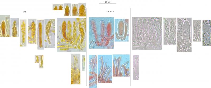
Asci: Inoperculate, 8-spored, croziers +, cylindrical to clavate, thick-walled, apex rounded to acute, one spore at apex, some spores horizontally oriented, spores often obliquely arranged, 1 – 2 (3) seriate.
Vital (presumed): Turgid, more clavate, apex more rounded, apical dome less pronounced (harder to see rings), pars sporifera ~50%.
Measured in tap water or IKI:
(30.6) 31.4 - 33.7 (34.2) × (6.3) 6.7 - 8 (8.4) µm,
Q = 4 - 4.5 (5.1), N = 7,
Me = 32.4 × 7.2 µm, Qe = 4.4.
Dead (presumed): Generally flaccid, often more narrowly-cylindrical, sometimes spores grouped towards base causing more lageniform shape, apex more acute and truncate, apical dome pronounced, some spores may be reversely oriented, pars sporifera ~80-90%.
Measured in KOH:
(28.2) 28.5 - 34.8 (36.1) × (4.1) 5.1 - 7.5 (8.3) µm,
Q = (4) 4.3 - 6.2 (7), N = 23,
Me = 31.8 × 6.1 µm, Qe = 5.3
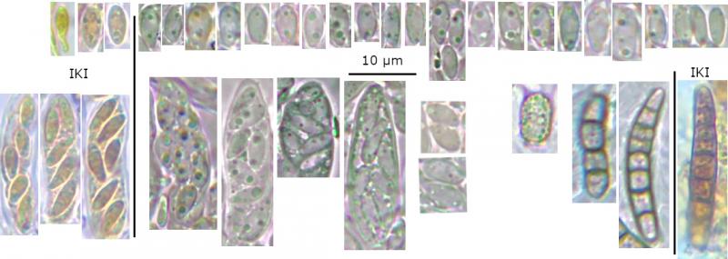
Spores: Ellipsoid-ovoid, often inequilateral in profile view, occasionally heteropolar?, usually multi-guttulate, 1 (- 2) medium-size and several smaller LBs towards each pole, OCI 1 - 3, no septa identified.
Vital spores in tap water squash mount, some measured in asci (agreeable with a few spores ejected from sections):
(4.7) 5.1 - 6 (6.3) × (2.4) 2.5 - 3.1 (3.5) µm,
Q = (1.6) 1.7 - 2.2 (2.4, N = 30,
Me = 5.6 × 2.8 µm, Qe = 2
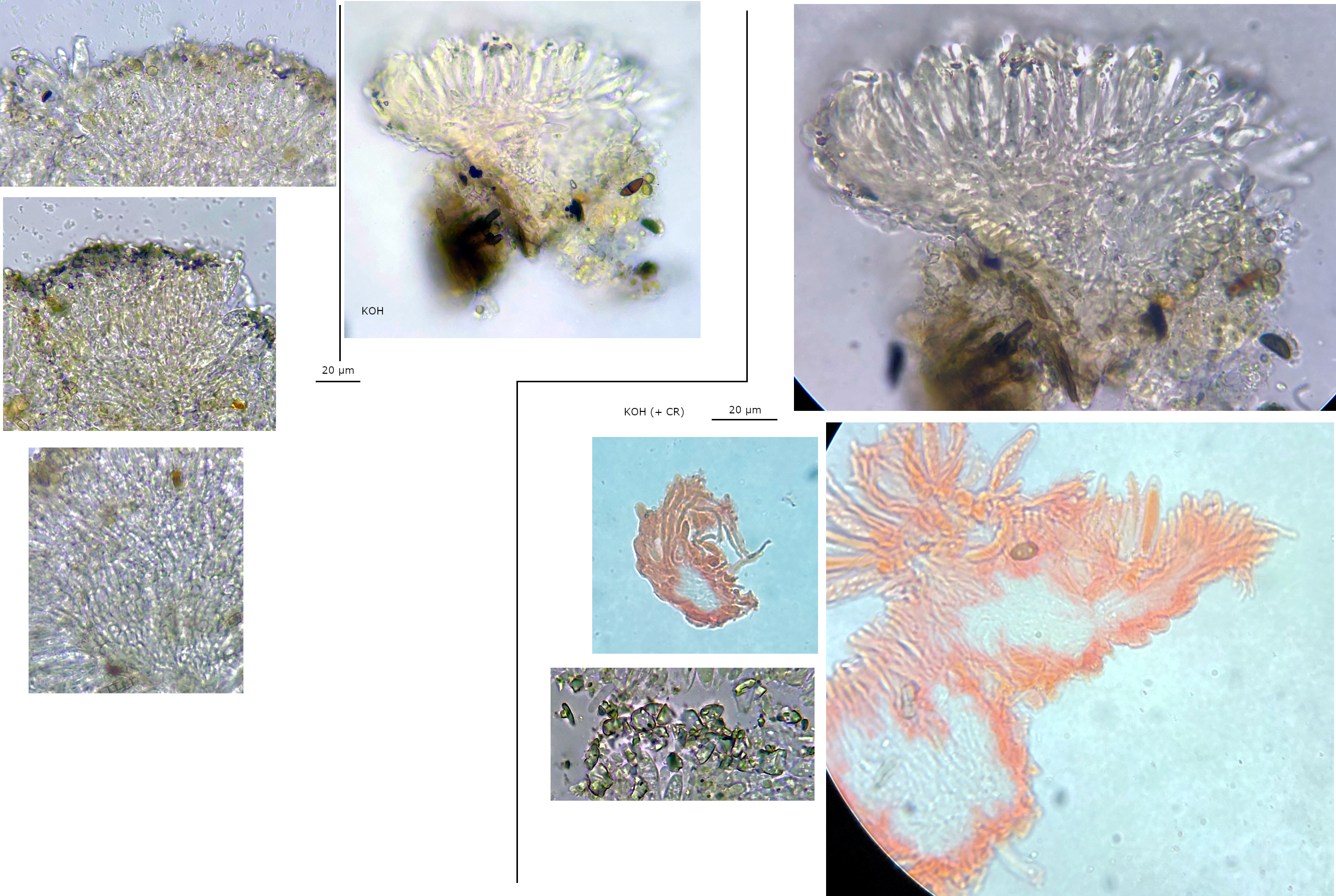
Subhymenium: Hyaline hyphae.
Marginal hairs: Hyaline, short, cylindrical (to lageniform?), crystallised, no ornamentation?, apex rounded, appear 2 – 3 septate.
Ectal excipulum: Hyaline, textura prismatica, possibly more textura angularis towards base.
Marginal cells: Elongated, one cell protruding, larger and more pyriform around the base, yellowish at base.
Exudate: Ochraceous-orange with crystals, noticeably orangish in concentration around margin, hyaline fragments in water mount, large hyaline crystals found in water mount may not be associated.
Medullary excipulum: Appears to be a small central column of hyaline hyphae that is textura intricata-porecta.
Anchoring hyphae: Some brownish hyphae around base.
Subiculum: None identified.
Anamorph: No direct associations identified.


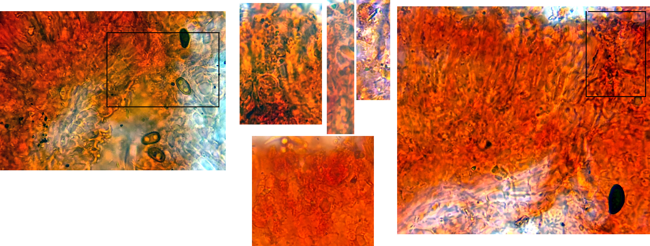
 Sections-0001.jpeg
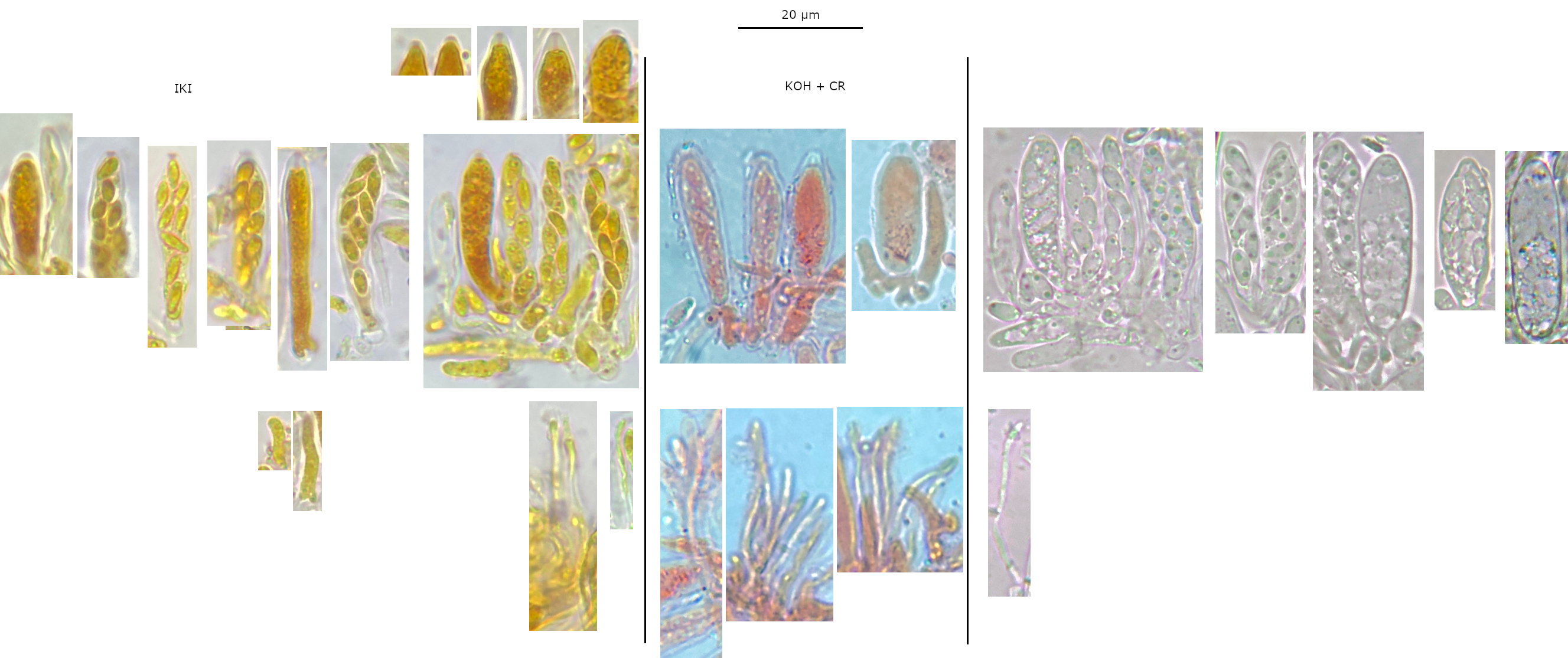 Hymenium-0001.jpeg
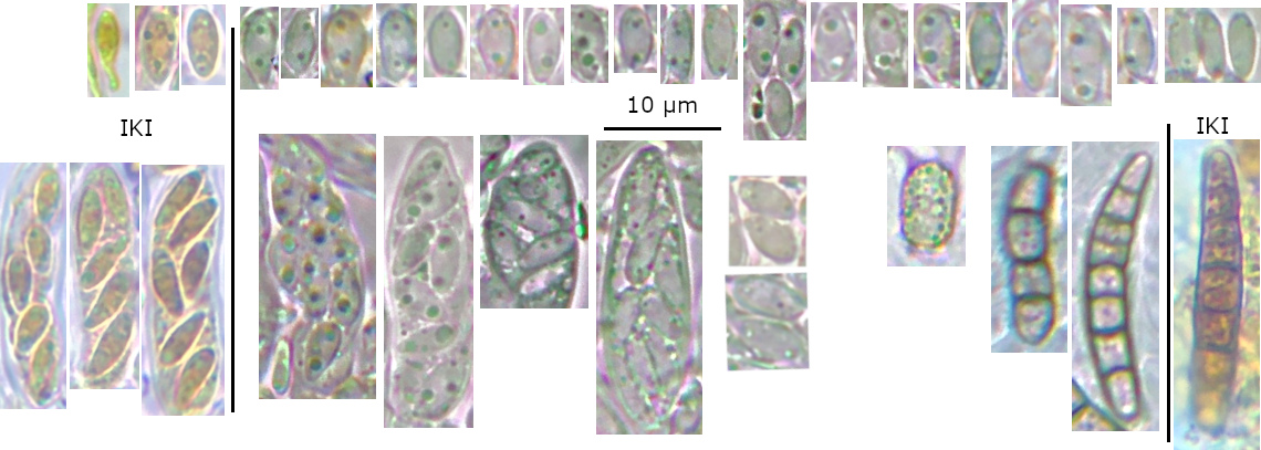 Spores-0001.jpeg
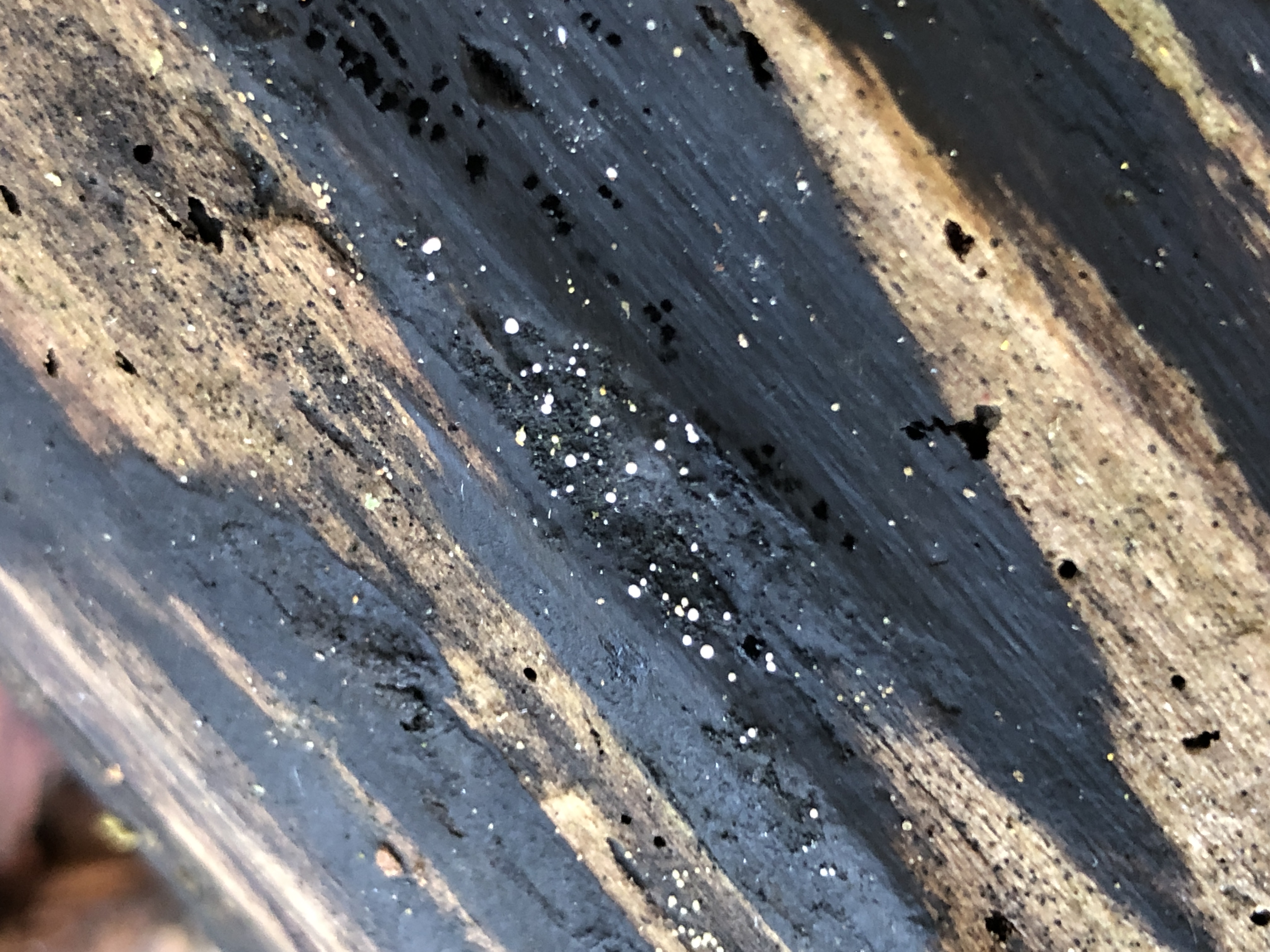 IMG-1834-0001.JPG
 Macro-1-0001.jpeg
 Macro-2-0001.jpeg
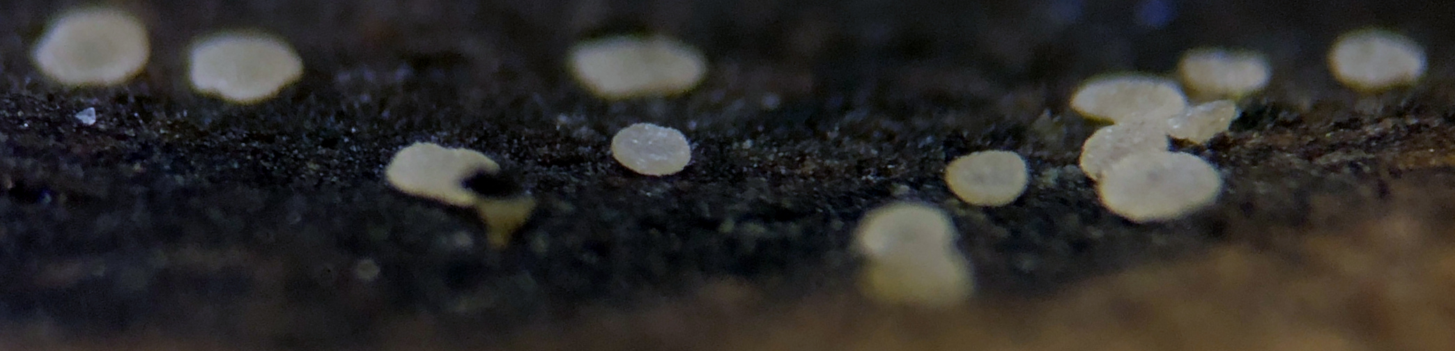 Macro-3-0001.jpeg
Look in direction Hyphodiscus, may be Hyphodiscus hyaloscyphoides.
You need better pictures from hairs.
https://drive.google.com/drive/u/0/folders/0B5SeyOEkxxZhazllLU1IemNVZlU?tid=0B5SeyOEkxxZhYVZub0N1aGY5YTg&resourcekey=0-6c-dufxw6WPl5FQxwsmRhQ
Greetings
Ingo W
I am glad you said that species, because it is what I got from the Quijada et al. key (ascus length, spore size, hemiamyloid, macro, habitat). I then convinced myself that the disc wasnt smooth enough for the genus/family and I didnt see any warty apical cells from hairs.
You can't see warty hairs in blurry pictures.
Show this sharp:
Greetings
Ingo W
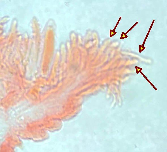
Unfortunately my sections got stuck together and the photos are rather disappointing. I may have another try to get better photos if I can face trying to handling the ascomata again.
 Hairs-0001.jpeg