
28-03-2016 21:27
 William Slosse
This species was growing on bark of Betula. Is m
28-03-2016 15:57
Cvenkel MiranConsider upper two to be red with no doubt. Spotte
28-03-2016 18:47
 Baeza Yajaira
Hello, Im looking for this paper Poelt J. Zur Sy


26-03-2016 21:50
 Bernard CLESSE
Je reste dans les Mniaecia avec cette belle espèc
24-03-2016 20:56
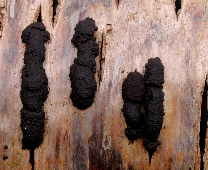
Thorben HülsewigHi there,today i found in a mixed forest (Quercus/
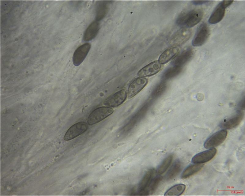
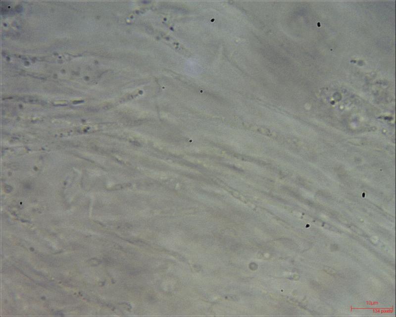
This species was growing on bark of Betula.
Is my determination correct?
Thx in advance,
W.

Hello,
definitely - what makes you hesitate?
best regards,
Andreas
Hi William,
oh, I didn't intend to be annoying ...
I was asking because I had some difficulties last summer in identifying H. mulitforme on Carpinus because the young stromata gave a brighter KOH reaction than described in pyrenomycetes.free.fr - but it was H. multiforme nevertheless. So it might be important to know that the young stoma yield a more orange reaction compared to the olivaceous-yellowish reaction of the mature stomata.
best regards,
Andreas