15-05-2016 13:19
 Rubén Martínez-Gil
Hola a todos. Subo unas fotos de un asco que enco
15-05-2016 02:06
 Alan Rockefeller
Hi - I recently sequenced one of my collections f
14-05-2016 10:57
Elisabeth StöckliBonjour, Trouvé au sol, sur feuille d' Andromeda
13-05-2016 21:07
 Enrique Rubio
Please, could you help me with this paper? Fungi
14-05-2016 15:00
 Blasco Rafael
Hola, les parece que sea la que propongo, las medi
20-08-2014 21:35
Marja PennanenHello folks,these beautyful ascos grow on a river
24-01-2011 01:21
Erwin GruberI posted the following entry at the item "sur prel

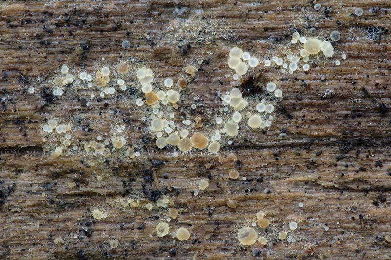
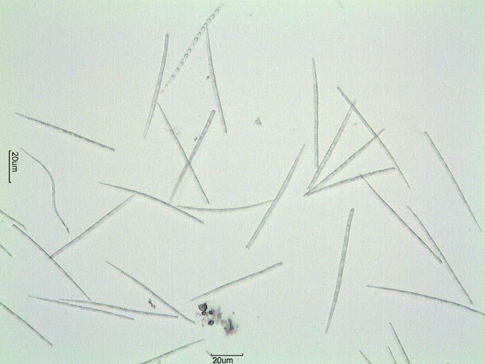
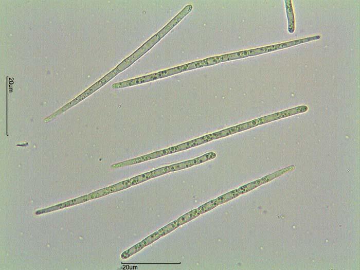
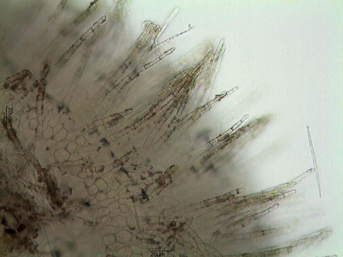
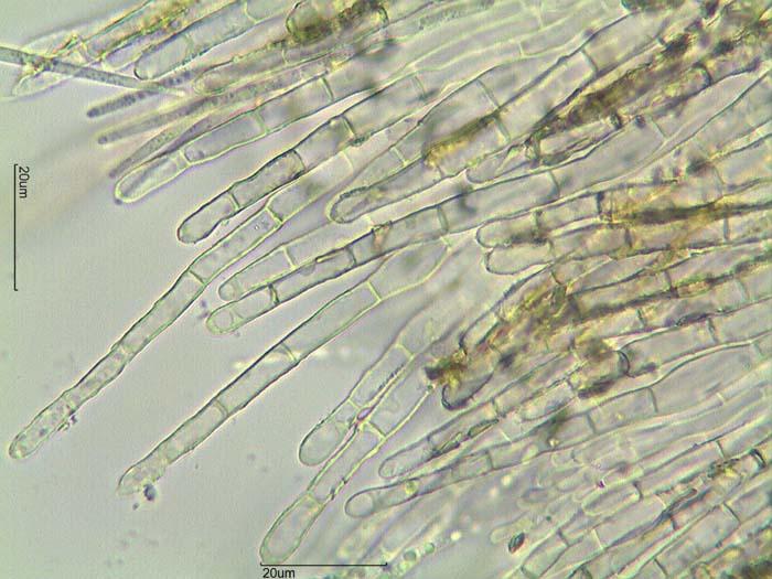
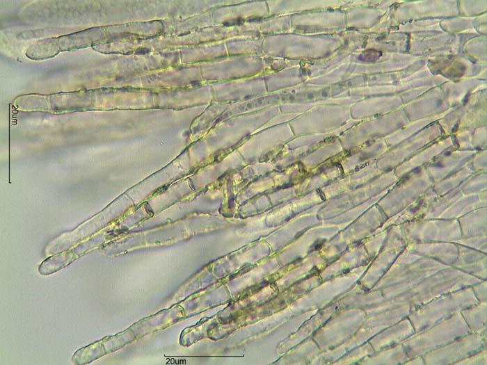
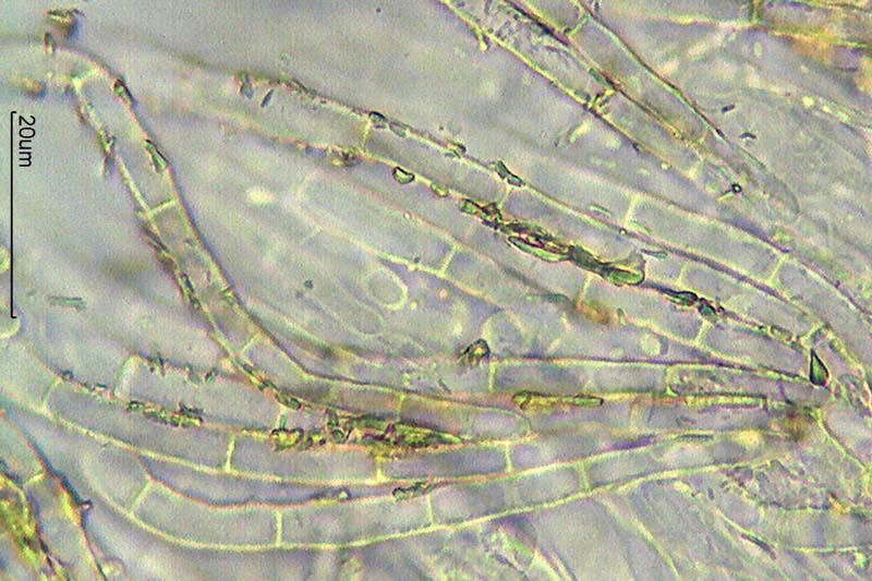
Hola a todos.
Subo unas fotos de un asco que encontramos ayer sobre madera semidescompuesta, creemos que de Quercus.
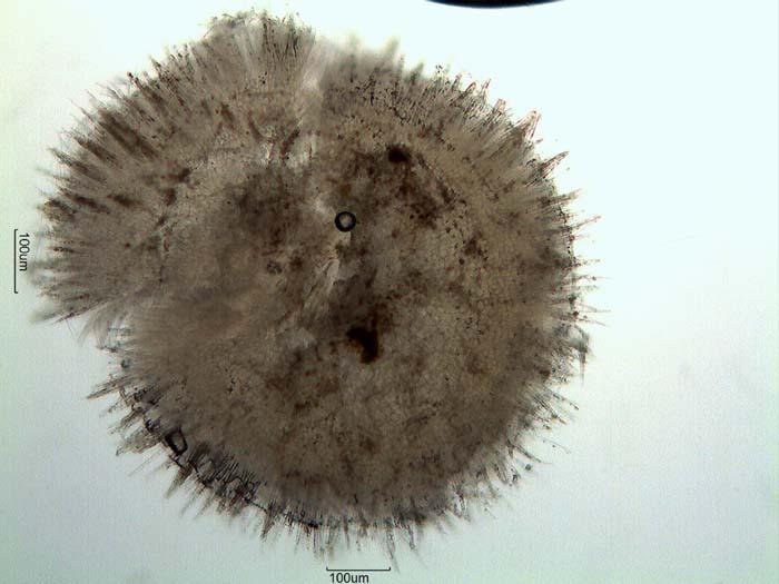
Apotecios de hasta 1 mm de diámetro.
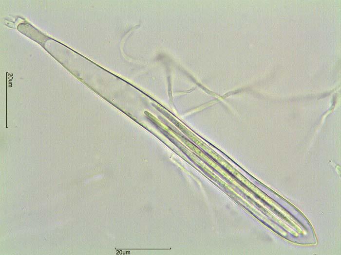
Ascas pleurorrincas, amiloides, de unas 130 x 12 micras.
Esporas de 60-80 x 2,2-2,8 micras con múltiples septos.
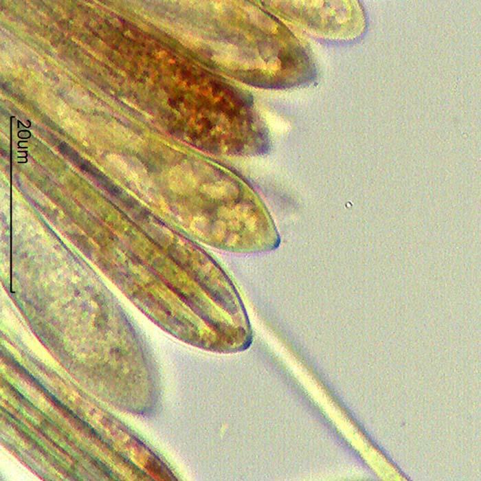
Pelos hialinos, con cristales, septados, de paredes delgadas y con ápice romo y base muy engrosada, de unas 50-110 x 4,5-7 micras.
Pensé en Arachnopeziza aurata, ¿Qué les parece?
Gracias por sus respuestas.
Rubén